. 2017 Sep 19;114(38):10220-10225.
doi: 10.1073/pnas.1706689114. Epub 2017 Sep 5.

Detection of immune responses after immunotherapy in glioblastoma using PET and MRI

Affiliations

Detection of immune responses after immunotherapy in glioblastoma using PET and MRI

Joseph P Antonios et al. Proc Natl Acad Sci U S A. .

Abstract

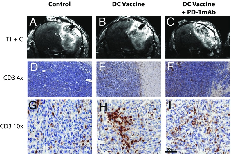
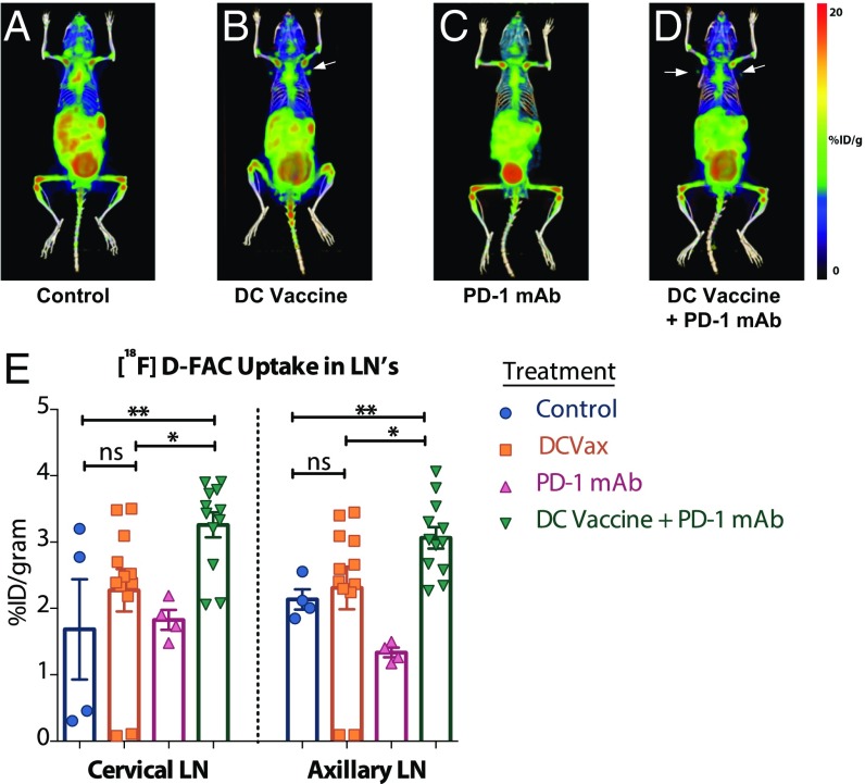
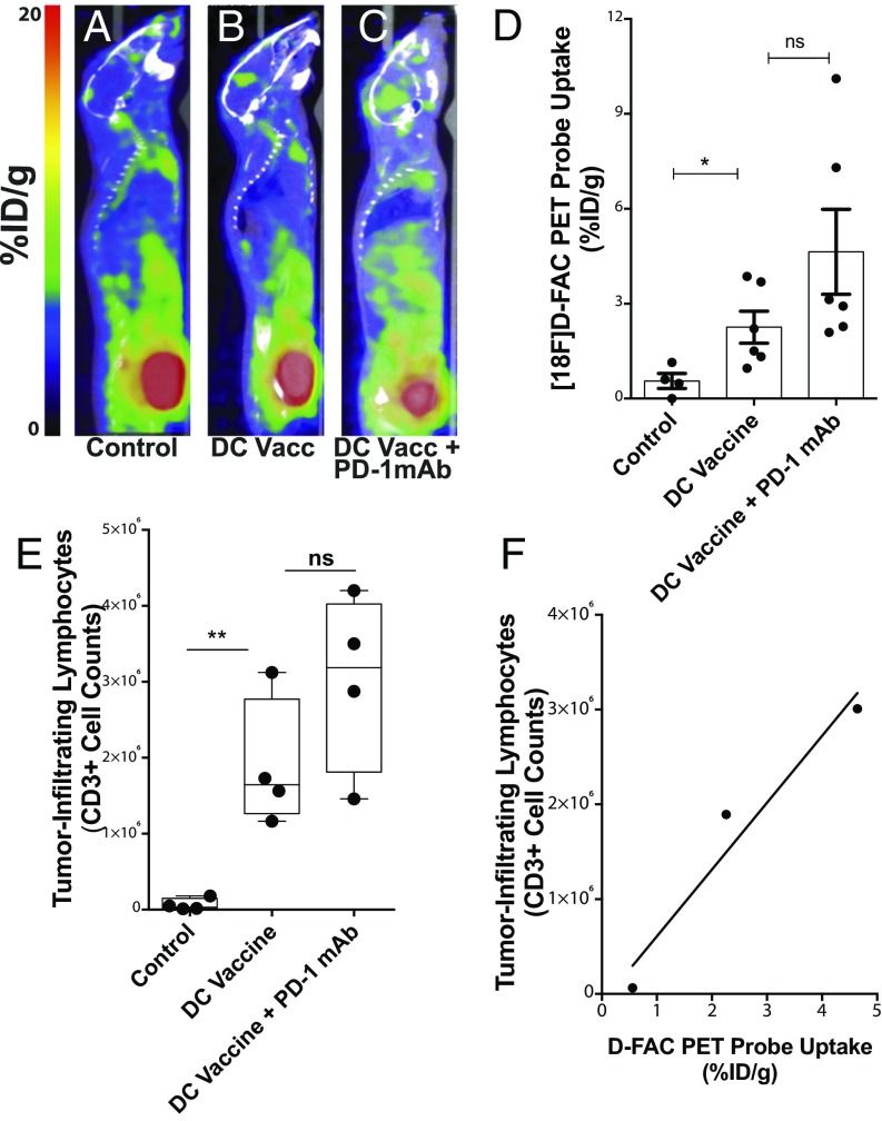
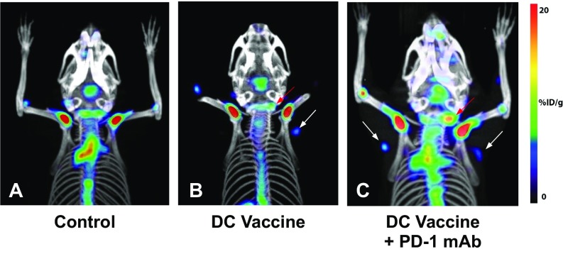
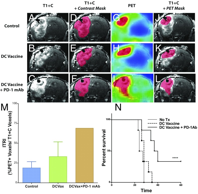
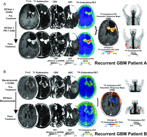
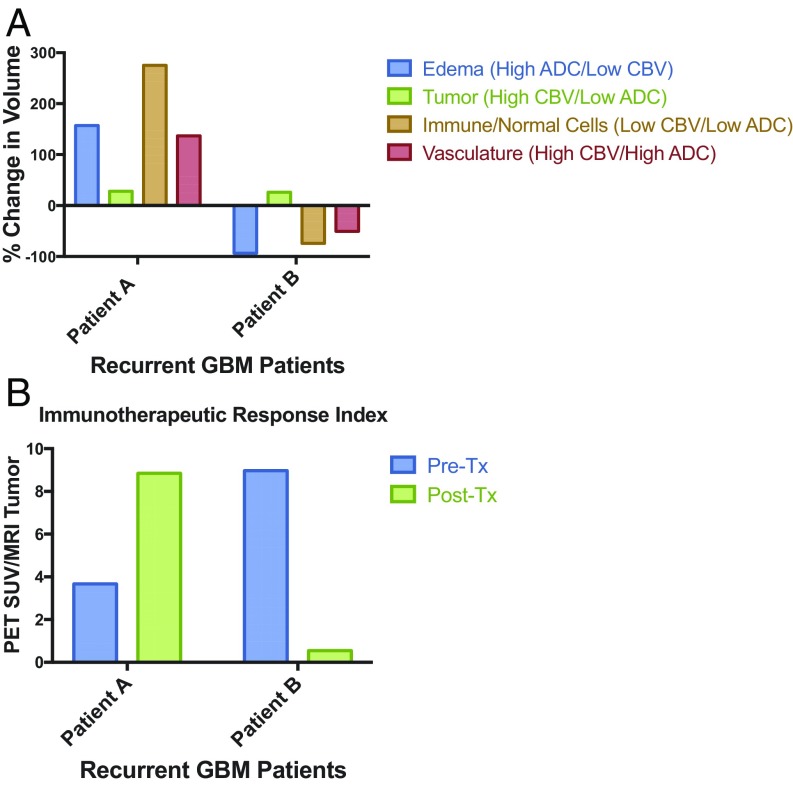
Contrast-enhanced MRI is typically used to follow treatment response and progression in patients with glioblastoma (GBM). However, differentiating tumor progression from pseudoprogression remains a clinical dilemma largely unmitigated by current advances in imaging techniques. Noninvasive imaging techniques capable of distinguishing these two conditions could play an important role in the clinical management of patients with GBM and other brain malignancies. We hypothesized that PET probes for deoxycytidine kinase (dCK) could be used to differentiate immune inflammatory responses from other sources of contrast-enhancement on MRI. Orthotopic malignant gliomas were established in syngeneic immunocompetent mice and then treated with dendritic cell (DC) vaccination and/or PD-1 mAb blockade. Mice were then imaged with [18F]-FAC PET/CT and MRI with i.v. contrast. The ratio of contrast enhancement on MRI to normalized PET probe uptake, which we term the immunotherapeutic response index, delineated specific regions of immune inflammatory activity. On postmortem examination, FACS-based enumeration of intracranial tumor-infiltrating lymphocytes directly correlated with quantitative [18F]-FAC PET probe uptake. Three patients with GBM undergoing treatment with tumor lysate-pulsed DC vaccination and PD-1 mAb blockade were also imaged before and after therapy using MRI and a clinical PET probe for dCK. Unlike in mice, [18F]-FAC is rapidly catabolized in humans; thus, we used another dCK PET probe, [18F]-clofarabine ([18F]-CFA), that may be more clinically relevant. Enhanced [18F]-CFA PET probe accumulation was identified in tumor and secondary lymphoid organs after immunotherapy. Our findings identify a noninvasive modality capable of imaging the host antitumor immune response against intracranial tumors.

Keywords: MRI; PET; checkpoint blockade; glioblastoma; immunotherapy.


Figures

Fig. 1.
Fig. 1.
Standard contrast-enhanced MRI cannot distinguish tumor growth from pseudoprogression in glioma-bearing mice. (AC) Representative coronal sections of T1-weighted MRI images following i.v. injection of contrast agent. (DI) 4× magnified (DF) and 10×- magnified (G–I) cross-sections of mice from control, DCVax, and DCVax + PD-1 mAb mice after immunohistochemical staining for CD3. Images were obtained from representative mice in experiments repeated multiple times; n = 4–6 mice per group.
Fig. 2.
Fig. 2.
DC vaccination with or without PD-1 mAb blockade results in elevated [18F]-FAC probe accumulation in lymph nodes. (AD) Representative 3D whole-body reconstructions of [18F]-FAC PET and CT imaging in control, DCVax, PD-1 mAb, and DCVax + PD-1 mAb treatment groups. (E) Regions of interest (ROIs) and quantification of probe uptake measured in the cervical and axillary lymph nodes in the treatment groups. *P < 0.05; **P < 0.005, unpaired t tests of the ROI data. n = 4–6/group. Measurements were repeated at least twice to verify the results.
Fig. 3.
Fig. 3.
DC vaccination with or without PD-1 mAb blockade results in elevated [18F]-FAC probe accumulation in gliomas and directly correlates with lymphocytic infiltrate. (AC) Representative whole-body cross-sections of PET-imaged mice injected with [18F]-FAC radioactive label for control, DCVax-treated, and DCVax + PD-1 mAb-treated mice. (D) [18F]-FAC PET probe uptake in tumor was significantly elevated in treated mice compared with untreated mice (*P < 0.05, **P < 0.01); n = 4–6/group. (E) Absolute counts of CD3+ tumor-infiltrating lymphocytes were significantly elevated in treated mice compared with untreated mice (**P < 0.01, ***P < 0.001); n = 4/group. (F) Correlation between the average PET probe uptake per group (D) and the average absolute infiltrating CD3+ lymphocyte count (E) per group quantified (R2 = 0.89) across treatment groups.
Fig. S1.
Fig. S1.
Increased [18F]-FAC probe accumulation in lymph nodes following treatment with DCVax ± PD-1 mAb blockade. (AC) Additional representative [18F]-FAC PET and CT images in control, DCVax, and DCVax + PD-1 mAb treatment groups highlighting cervical and axillary lymph node tracer uptake.
Fig. 4.
Fig. 4.
The ITRI predicts survival outcome in glioma-bearing mice following immunotherapy. (AC) Representative coronal T1-weighted MRI sections of untreated control, DCVax-treated, and DCVax + PD-1 mAb-treated mice. (DF) Representative contrast subtraction maps (red; contrast mask) overlaid onto T1-weighted MRI images with contrast. (GI) Representative coronal [18F]-FAC PET images of untreated control and DCVax- and DCVax + PD-1 mAb-treated mice. (JL) Representative threshold PET subtraction maps (red; PET mask) overlaid onto T1-weighted MRI images with contrast. (M) The ITRI (% PET voxels/T1+C voxels) calculated for each treatment group. n = 1–4 mice/group. Calculations were performed twice, with similar findings. (N) Survival of intracranial GBM-bearing untreated control (no Tx), DCVax-treated, and DCVax + PD-1 mAb-treated mice (****P < 0.0001); n = 6/group.
Fig. 5.
Fig. 5.
The combination of [18F]-CFA PET and advanced MRI can help distinguish tumor progression from inflammation in patients with GBM treated with DC vaccination and PD-1 mAb blockade. Postcontrast T1-weighted, T1-subtraction, relative CBV, ADC, [18F]-FAC PET + MRI fusion, and whole-body maximum-intensity projection images of [18F]-CFA from two patients (A, patient A; B, patient B) with recurrent GBM before (Top) and after (Bottom) immunotherapy.
Fig. 6.
Fig. 6.
Quantitative estimation of intratumoral immune responses using combined multiparametric MRI/PET. (A) Quantification of edema, tumor, immune/normal cells, and vasculature from perfusion (CBV) and diffusion (ADC)-weighted MRI. (B) Ratio of [18F]-CFA PET standard uptake value divided by the tumor (high CBV/low ADC) volumes for patients A and B.

References

    1. Antonios JP, et al. PD-1 blockade enhances the vaccination-induced immune response in glioma. JCI Insight. 2016;1:e87059. - PMC - PubMed
    1. Liau LM, et al. Dendritic cell vaccination in glioblastoma patients induces systemic and intracranial T-cell responses modulated by the local central nervous system tumor microenvironment. Clin Cancer Res. 2005;11:5515–5525. - PubMed
    1. Prins RM, et al. Gene expression profile correlates with T-cell infiltration and relative survival in glioblastoma patients vaccinated with dendritic cell immunotherapy. Clin Cancer Res. 2011;17:1603–1615. - PMC - PubMed
    1. Prins RM, et al. Comparison of glioma-associated antigen peptide-loaded versus autologous tumor lysate-loaded dendritic cell vaccination in malignant glioma patients. J Immunother. 2013;36:152–157. - PMC - PubMed
    1. Hsu M, et al. TCR Sequencing can identify and track glioma-infiltrating T cells after DC vaccination. Cancer Immunol Res. 2016;4:412–418. - PMC - PubMed

Publication types