. 2017 Sep 5;8(1):427.
doi: 10.1038/s41467-017-00573-w.

Signalling strength determines proapoptotic functions of STING

Affiliations

Signalling strength determines proapoptotic functions of STING

Muhammet F Gulen et al. Nat Commun. .

Abstract

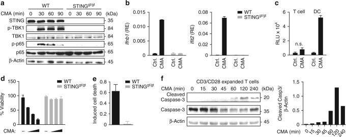
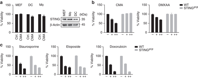
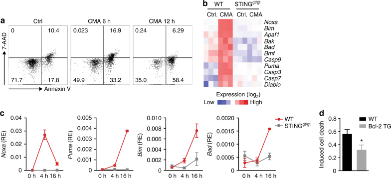
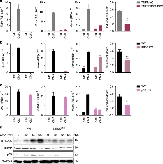
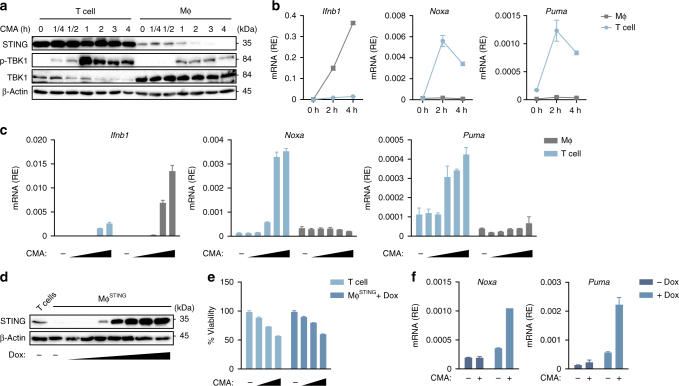
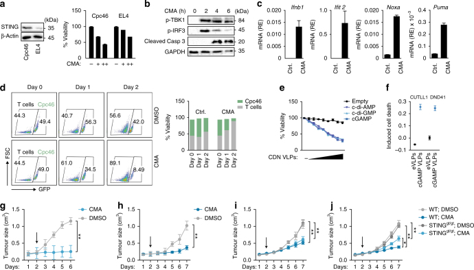
Mammalian cells use cytosolic nucleic acid receptors to detect pathogens and other stress signals. In innate immune cells the presence of cytosolic DNA is sensed by the cGAS-STING signalling pathway, which initiates a gene expression programme linked to cellular activation and cytokine production. Whether the outcome of the STING response varies between distinct cell types remains largely unknown. Here we show that T cells exhibit an intensified STING response, which leads to the expression of a distinct set of genes and results in the induction of apoptosis. Of note, this proapoptotic STING response is still functional in cancerous T cells and delivery of small molecule STING agonists prevents in vivo growth of T-cell-derived tumours independent of its adjuvant activity. Our results demonstrate how the magnitude of STING signalling can shape distinct effector responses, which may permit for cell type-adjusted behaviours towards endogenous or exogenous insults.The cGAS/STING signalling pathway is responsible for sensing intracellular DNA and activating downstream inflammatory genes. Here the authors show mouse primary T cells and T leukaemia are hyperresponsive to STING agonist, and this strong STING signalling is associated with apoptosis induction.


Conflict of interest statement

The authors declare no competing financial interests.

Figures

Fig. 1
Fig. 1
STING activation induces apoptosis in T cells. a CD4+ T cells from wild-type (WT) or STINGgt/gt mice were treated with DMSO or 10-carboxymethyl-9-acridanone (CMA) for the indicated times and phosphorylation of TBK1 and NF-κB (p65) was assessed by immunoblot. b CD4+ T cells from WT or STINGgt/gt mice were treated with DMSO or CMA for 4 h and abundance of mRNAs of the indicated genes was measured by RT-qPCR. c Supernatants from CD4+ T cells or dendritic cells (DC) treated with DMSO or CMA were collected after 16 h. A bioassay was used to measure the production of type I IFNs (RLU relative light units). d, e CD4+ T cells from WT and STINGgt/gt mice were treated with DMSO or CMA. After 16 h cell viability d and percentage of apoptotic cells e, assessed by Annexin V staining, were determined. f WT CD4+ T cells were stimulated with CMA for the indicated times and caspases-3 cleavage was assessed by immunoblot. Quantification of cleaved Caspase-3 relative to β-Actin levels is shown on the right panel. Data are representative of n = 3 (a, f) independent experiments or mean and s.d. of technical replicates (n = 2) of one representative experiment out of n = 3 (b, d, e) or n = 2 (c) independent experiments are shown. Unprocessed original blots are shown in Supplementary Fig. 9.
Fig. 2
Fig. 2
STING activation induces apoptosis in a cell type-specific manner. a Left: Primary mouse embryonic fibroblasts (MEF), bone marrow-derived macrophages (MΦ) and bone marrow-derived DCs (DC) were treated with DMSO or 10-carboxymethyl-9-acridanone (CMA) and 16 h later cell viability was determined. Right: STING expression in distinct cell types was analysed by immunoblot. b, c CD4+ T cells from wild-type (WT) and STINGgt/gt mice were left untreated or stimulated with CMA (0.125 mg/ml, 0.25 mg/ml), DMXAA (10 µg/ml, 100 µg/ml), staurosporine (100 nM, 500 nM), etoposide (1 µg/ml, 5 µg/ml) or doxorubicin (0.4 µg/ml, 1 µg/ml) overnight. Cell viability was assessed by CellTiter Blue assay. Representative results from n = 3 independent experiments (a; right panel) or mean and s.d. of technical replicates of one representative experiment out of n = 3 experiments are shown ac. Unprocessed original blots are shown in Supplementary Fig. 9
Fig. 3
Fig. 3
BH3-only proteins are involved in STING-triggered T cell apoptosis. a Flow cytometric analysis of apoptosis of CD4+ T cells left untreated or treated with 10-carboxymethyl-9-acridanone (CMA) for 6 h and 12 h, assessed by Annexin V and 7-AAD staining. Numbers in quadrants present percentages. b Heat-map of RNA-sequencing analysis. Genes related to cell intrinsic apoptosis with statistically significant increase in CMA-treated over DMSO-treated CD4+ T cells are shown (n = 3 biological replicates; P < 0.05; Student’s t-test). c T cells from wild-type (WT) or STINGgt/gt mice were untreated or stimulated with CMA for 4 h and 16 h. Abundance of mRNAs for the indicated genes was quantified by RT-qPCR. d CD4+ T cells of the genotypes indicated were treated with DMSO or CMA. After 16 h the percentage of apoptotic cells was assessed by Annexin V and 7-AAD staining. Data are representative of n = 3 independent experiments (a) or mean and s.d. of technical replicates of one representative experiment out of n = 3 (c) or n = 2 (d) independent experiments are shown. *P < 0.05; (Student’s t-test)
Fig. 4
Fig. 4
Cooperation of IRF-3 and p53 for the STING-mediated proapoptotic transcriptional programme. ac CD4+ T cells of the indicated genotypes were treated with DMSO or 10-carboxymethyl-9-acridanone (CMA). After 16 h the percentage of apoptotic cells was assessed by Annexin V and 7-AAD staining (right) or upregulation of depicted genes was determined by RT-qPCR (left). d CD4+ T cells from wild-type (WT) or STINGgt/gt mice were stimulated with CMA for the indicated times and p53, MDM2 and phosphorylation of Histone H2A.X were assessed by immunoblot. Data are representative of n = 2 d independent experiments or mean and s.d. of technical replicates of one representative experiment out of n = 3 independent experiments are shown ac. *P < 0.05 and **P < 0.01 (Student’s t-test). Unprocessed original blots are shown in Supplementary Fig. 9
Fig. 5
Fig. 5
Intensified STING signalling leads to apoptosis in T cells. a Wild-type (WT) CD4+ T cells and bone marrow-derived macrophages (MΦ) were left untreated or stimulated with 10-carboxymethyl-9-acridanone (CMA) for the indicated time points and immunoblot analysis was performed with indicated antibodies. b WT CD4+ T cells and macrophages were stimulated with CMA for 2 h and 4 h, followed by quantification of abundance of mRNAs for the indicated genes by RT-qPCR. c WT CD4+ T cells and macrophages were stimulated with increasing doses of CMA (31.25–250 μg/ml) and upregulation of depicted genes was determined by RT-qPCR. d Macrophages transduced with a doxycycline-inducible murine STING construct (MΦSTING) were treated with doxycycline and STING expression was determined by immunoblot. e WT CD4+ T cells and MΦSTING were treated with CMA and cell viability was assessed by CellTiter Blue assay. fSTING in the presence or absence of doxycycline were treated with CMA and proapoptotic gene expression was assessed by RT-qPCR. Data are representative of three independent experiments a, d or mean and s.d. of technical replicates of one representative experiment out of n = 3 independent experiments are shown b, c, e, f. Unprocessed original blots are shown in Supplementary Fig. 9
Fig. 6
Fig. 6
Pharmacological hyperactivation of STING promotes apoptosis of T-cell-derived malignant cells. a Murine T-ALL cells (Cpc46) and lymphoma cells (EL4) were incubated with 10-carboxymethyl-9-acridanone (CMA) for 16 h and cell viability was assessed by CellTiter Blue assay. b Cpc46 cells were treated with DMSO (upper panel) or CMA (lower panel) and phosphorylation of TBK1, IRF-3 and cleavage of caspase-3 was assessed at indicated times by immunoblot. c Cpc46 cells were treated with DMSO or CMA and the induction of indicated genes was quantified by RT-qPCR after 4 h. d Primary CD4+ T cells (in the presence of CD3 and CD28 activation) and Cpc46 cells (x axis, GFP+ cells) were cultured in a 1:1 ratio and treated with DMSO (upper panel) or CMA (lower panel). After 1 day and 2 days percentages of GFP+ cells (Cpc46 cells) vs. GFP primary T cells were determined by FACS. e Cpc46 cells were incubated with empty viral particles (eVLPs) or VLPs containing cGAMP (cGAMP VLPs), c-di-AMP or c-di-GMP for 16 h and cell viability was assessed by CellTiter Blue assay. f CUTLL1 cells and DND41 cells were left untreated or incubated with eVLPs or cGAMP VLPs and percentages of apoptotic cells were measured by FACS after 16 h. gj Subcutaneous in vivo growth of Cpc46 cells (g) and EL4 cells (h) in Rag2−/−γc−/− mice or Cpc46 cells (i) and EL4 cells (j) in wild-type (WT) or STINGgt/gt mice that were treated with or without (Ctrl.; treatment with diluent only) CMA (500 µg), daily after tumour size reached up to 200 mm3. (Cpc46, n = 3 mice per treatment group; EL4, n = 5 mice per treatment group). Tumour size was measured daily. Arrow indicates starting point of the treatment. Data are representative of n = 3 (a; left) or n = 2 (b) independent experiments or mean and s.d. of one representative experiment out of n = 3 (a, cf) or mean and s.d of n = 3 (Cpc46 cells) or n = 5 (EL4 cells) mice per treatment group are shown gj. **P < 0.01 (two-way ANOVA). Unprocessed original blots are shown in Supplementary Fig. 9

References

    1. Wu J, Chen ZJ. Innate immune sensing and signaling of cytosolic nucleic acids. Annu. Rev. Immunol. 2014;32:461–488. doi: 10.1146/annurev-immunol-032713-120156. - DOI - PubMed
    1. Sun L, Wu J, Du F, Chen X, Chen ZJ. Cyclic GMP-AMP synthase is a cytosolic DNA sensor that activates the type I interferon pathway. Science. 2013;339:786–791. doi: 10.1126/science.1232458. - DOI - PMC - PubMed
    1. Wu J, et al. Cyclic GMP-AMP is an endogenous second messenger in innate immune signaling by cytosolic DNA. Science. 2013;339:826–830. doi: 10.1126/science.1229963. - DOI - PMC - PubMed
    1. Gao P, et al. Cyclic [G(2ʹ,5ʹ)pA(3ʹ,5ʹ)p] is the metazoan second messenger produced by DNA-activated cyclic GMP-AMP synthase. Cell. 2013;153:1094–1107. doi: 10.1016/j.cell.2013.04.046. - DOI - PMC - PubMed
    1. Ablasser A, et al. cGAS produces a 2ʹ-5ʹ-linked cyclic dinucleotide second messenger that activates STING. Nature. 2013;498:380–384. doi: 10.1038/nature12306. - DOI - PMC - PubMed

Publication types