. 2017 Sep 5;7(1):10565.
doi: 10.1038/s41598-017-10999-3.

FOXO1 deletion in keratinocytes improves diabetic wound healing through MMP9 regulation

Affiliations

FOXO1 deletion in keratinocytes improves diabetic wound healing through MMP9 regulation

Chenying Zhang et al. Sci Rep. .

Abstract

Keratinocyte migration is a key aspect of re-epithelialization during wound healing. Matrix metalloproteinase 9 (MMP9) contributes to this process and deficiencies in the MMP9 lead to impaired healing. Inappropriate expression of MMP9 also contributes to impaired re-epithelialization. Previously we demonstrated that FOXO1 was activated in wound healing but to higher levels in diabetic wounds. To address mechanisms of impaired re-epithelialization we examined MMP9 expression in vivo in full thickness dermal scalp wounds created in experimental K14.Cre + .Foxo1 L/L mice with lineage-specific Cre recombinase deletion of floxed FOXO1 and compared the results to control littermates. MMP9 was induced during wound healing but at a significantly higher level in diabetic compared to normal wounds. FOXO1 deletion substantially blocked this increase. By chromatin immunoprecipitation FOXO1 was shown to bind to the MMP9 promoter, FOXO1 overexpression increased MMP9 transcriptional activity and increased MMP9 expression stimulated by high glucose was blocked by FOXO1 deletion or FOXO1 knockdown. We also show for the first time that high glucose impairs keratinocyte migration by inducing high levels of MMP9 expression and establish that it involves FOXO1. Thus, FOXO1 drives high levels of MMP9 expression in diabetic wound healing, which represents a novel mechanism for impaired re-epithelization in diabetic wounds.


Conflict of interest statement

The authors declare that they have no competing interests.

Figures

Figure 1
Figure 1
Keratinocyte-specific FOXO1 deletion reduces MMP9 expression during wound healing. Dermal wounds were created in normoglycemic (NG) and diabetic experimental (K14.Cre+.Foxo1L/L) and littermate control (K14.Cre. Foxo1L/L) mice. (A) Representative images of MMP9 immunofluorescent staining with the MMP9 specific antibody on day 4 wounds. Scale bar, 100 µm. EP = epidermis, CT = connective tissues, white dashed lines demarcate the epidermis from the dermis. (B) MMP9 immunofluorescence analyses for day 4 wounds. (C) MMP9 immunofluorescence analyses for day 7 wounds. Each in vivo value is the mean ± SEM for n = 5–8 mice per group. *P < 0.05 vs. matched Cre group, # P < 0.05 vs. matched NG Cre- group.
Figure 2
Figure 2
FOXO1 deletion in keratinocytes reduces MMP9 expression in vitro. Primary murine keratinocytes (A-C) from K14.Cre.Foxo1 L/L or K14.Cre+.Foxo1 L/L mice and primary normal human epidermal keratinocytes (NHEK) cells (DF) were incubated in low glucose (LG) or in high glucose (HG) media. (A and B) MMP9 immunofluorescence analyses of murine keratinocyte immune-positive cells (A) and MMP9 fluorescence intensity (B). (C) MMP9 ELISA analyses of conditioned media from primary murine K14.Cre.Foxo1 L/L or K14.Cre+.Foxo1 L/L keratinocytes in LG or HG condition. (D and E) qRT-PCR analysis of MMP9 (D) and TIMP1 (E) mRNA levels. (F) The ratio of MMP9 to TIMP1 mRNA levels. Each data represents the mean ± SEM of 3 independent experiments. *P < 0.05 vs. matched Cre-, # P < 0.05 vs. LG Cre group, **P < 0.05 vs. matched scrambled siRNA, + P < 0.05 vs. matched LG group.
Figure 3
Figure 3
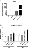
FOXO1 regulates the protein level of MMP9 in keratinocytes. (A) FOXO1 mRNA analyses in NHEK cells after transfection with control plasmid, wild-type FOXO1 plasmid (FOXO1-WT), or constitutively active FOXO1 plasmid (FOXO1-AAA) in LG media. (B) MMP9 fluorescence intensity in NHEK cells after transfection with control plasmid, wild-type FOXO1 plasmid (FOXO1-WT), or constitutively active FOXO1 plasmid (FOXO1-AAA) in LG or HG media. *P < 0.05 vs. matched control plasmid.
Figure 4
Figure 4
Inhibition of MMP9 rescues keratinocyte migration in a FOXO1 dependent manner in high glucose but not low glucose conditions. (A and B) Migration was measured by transwell assay for NHEK cells in the presence of conditioned media (CM) with or without indicated dose of active MMP9 protein in LG (A) or HG (B) conditions. (C) Migration was measured by transwell assay for primary murine keratinocytes isolated from K14.Cre.Foxo1 L/L or K14.Cre+.Foxo1 L/L mice in LG condition with CM. (D) Migration was measured by transwell assay for primary murine keratinocytes isolated from K14.Cre.Foxo1 L/L or K14.Cre+.Foxo1 L/L mice in HG condition with CM. Data represent the mean ± SEM of 3 independent experiments. *P < 0.05 vs. matched control group, **P < 0.05 vs. matched CM control group, + P < 0.05 vs. matched Cre- group.
Figure 5
Figure 5
FOXO1 is recruited to the MMP9 promoter and transactivates its expression. (A) ChIP assays for the binding of FOXO1 to the MMP9 promoter in NHEK cells in LG and HG conditions. ChIP-enriched DNA was quantified by qRT-PCR and values expressed as a percentage of input DNA. (B) NHEK cells were co-transfected with a control pcDNA3.1 vector or a vector that expresses constitutively active FOXO1 (FOXO1-AAA), control siRNA, or siRNA specific to FOXO1, together with a MMP9 reporter plasmid. Renilla luciferase reporter was used as an internal control. Luciferase activity was measured 36 h after transfection. Data represent the mean ± SEM of 3 independent experiments. *P < 0.05 vs. matched IgG control, # P < 0.05 vs. matched LG group, **P < 0.05 vs. matched control plasmid, ## P < 0.05 vs. matched scrambled siRNA.

References

    1. Hu SC, Lan CE. High-glucose environment disturbs the physiologic functions of keratinocytes: Focusing on diabetic wound healing. J DERMATOL SCI. 2016;84:121–127. doi: 10.1016/j.jdermsci.2016.07.008. - DOI - PubMed
    1. Giacco F, Brownlee M. Oxidative stress and diabetic complications. CIRC RES. 2010;107:1058–1070. doi: 10.1161/CIRCRESAHA.110.223545. - DOI - PMC - PubMed
    1. Liu Y, et al. Increased matrix metalloproteinase-9 predicts poor wound healing in diabetic foot ulcers: Response to Muller et al. DIABETES CARE. 2009;32 doi: 10.2337/dc09-1394. - DOI - PubMed
    1. Falanga V. Wound healing and its impairment in the diabetic foot. LANCET. 2005;366:1736–1743. doi: 10.1016/S0140-6736(05)67700-8. - DOI - PubMed
    1. Ravanti L, Kahari VM. Matrix metalloproteinases in wound repair (review) INT J MOL MED. 2000;6:391–407. - PubMed

Publication types