. 2017 Sep 5;7(1):10433.
doi: 10.1038/s41598-017-10438-3.

Hyperactive TGF-β Signaling in Smooth Muscle Cells Exposed to HIV-protein(s) and Cocaine: Role in Pulmonary Vasculopathy

Affiliations

Hyperactive TGF-β Signaling in Smooth Muscle Cells Exposed to HIV-protein(s) and Cocaine: Role in Pulmonary Vasculopathy

Pranjali Dalvi et al. Sci Rep. .

Abstract

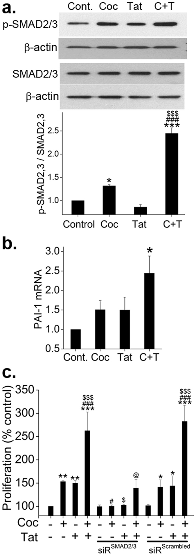
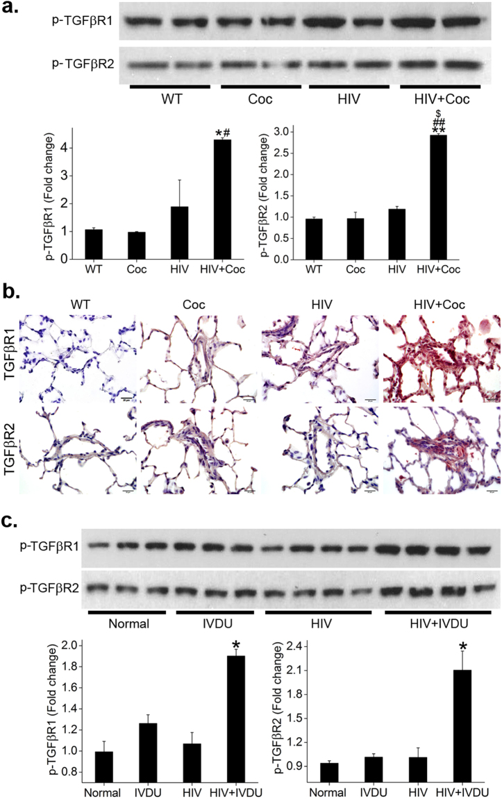
We earlier demonstrated synergistic increase in the proliferation of pulmonary smooth muscle cells on exposure to HIV-proteins and/or cocaine due to severe down-modulation of bone morphogenetic protein receptor (BMPR) axis: the anti-proliferative arm of TGF-β super family of receptors. Here, now we demonstrate the effect of HIV-Tat and cocaine on the proliferative TGF-β signaling cascade. We observed a significant increase in the secretion of TGF-β1 ligand along with enhanced protein expression of TGFβ Receptor (TGFβR)-1, TGFβR-2 and phosphorylated SMAD2/3 in human pulmonary arterial smooth muscle cells on treatment with cocaine and Tat. Further, we noticed an increase in the levels of p-TAK1 complexed with TGFβR-2. Concomitant to this a significant increase in the activation of TAK1-mediated, SMAD-independent downstream signaling molecules: p-MKK4 and p-JNK was observed. However, activation of MKK3/6-p38MAPK, another axis downstream of TAK1 was found to be reduced due to attenuation in the protein levels of BMPR2. Both SMAD and non-SMAD dependent TGFβR cascades were found to contribute to hyper-proliferation. Finally the increase in the levels of phosphorylated TGFβR1 and TGFβR2 on exposure to HIV-proteins and cocaine was confirmed in pulmonary smooth muscle cells from cocaine injected HIV-transgenic rats and in total lung extracts from HIV infected cocaine and/or opioid users.


Conflict of interest statement

The authors declare that they have no competing interests.

Figures

Figure 1
Figure 1
Cocaine and Tat induced increase in TGFβ ligand and receptor expression stimulates HPASMC proliferation. Quiescent HPASMCs were treated with cocaine (Coc) 1 µM and/or Tat (25ng/ml). (a) Cell supernatants collected from 1 to 12 days post-treatment were analyzed for TGFβ1 by ELISA. All values are mean ± SD of 3 independent experiments done in duplicates. *p ≤ 0.05 **p ≤ 0.01, ***p ≤ 0.001 compared to untreated control (Cont.), $p ≤ 0.001 vs. Tat; #p ≤ 0.001 vs. Coc. (b) Cells treated for 2, 3 or 6 days were lysed using RIPA buffer followed by western blot for TGFβR1 and 2. The graphs represent average densitometry of 3 independent experiments. (c) HPASMCs (3 × 103/well) were seeded in 96 well plate, 48 h after which the medium was replaced with 0.1% serum containing medium followed by cocaine and/or Tat treatment for 2 days in presence or absence of TGFβR1 inhibitor: SB431542. MTS cell proliferation assay was then conducted. All values are mean ± SD of at least three independent experiments done in triplicates. *p ≤ 0.05 **p ≤ 0.01, ***p ≤ 0.001 compared to untreated control (Cont.), $p ≤ 0.05, $$p ≤ 0.01, $$$p ≤ 0.001 compared to Tat; ##p ≤ 0.01, ###p ≤ 0.001 compared to Coc, @p ≤ 0.001 compared to cocaine and Tat combined treatment (C + T).
Figure 2
Figure 2
Augmented SMAD dependent TGFβ signaling in HPASMCs on exposure to combined treatment of cocaine and HIV-Tat. Quiescent HPASMCs were treated with cocaine 1 µM and/or Tat (25 ng/ml) for 2 days for western blot and RNA analysis. (a) Representative images of the Western blots probed using total and phosphorylated (p) - SMAD2/3. Graph represents average densitometry of 3 independent experiments. (b) Real time RT-PCR analysis of PAI1 mRNA expression. (c) HPASMC (3 × 103/well) were seeded in 96 well plate followed by either transfection with siRNASMAD2/3 or siRNAscrambled. After 48 h, the medium was replaced with 0.1% serum containing medium followed by cocaine and/or Tat treatment for 2 days. MTS cell proliferation assay was then conducted. All values are mean ± SD of at least three independent experiments performed in triplicates. *p ≤ 0.05 **p ≤ 0.01, ***p ≤ 0.001 compared to untreated control (Cont.), $p ≤ 0.05, $$$p ≤ 0.001 compared to Tat; #p ≤ 0.05, ###p ≤ 0.001 compared to cocaine (Coc), @p ≤ 0.001 compared to C + T.
Figure 3
Figure 3
Cocaine-Tat mediated increase in TGFβR2 and TAK1 complex formation in hyperproliferative HPASMCs. Protein was extracted from quiescent HPASMCs treated with cocaine and/or Tat for 2 days followed by (a) Western blot for p-TAK1, TAK1 or (b) Immunoprecipitation (IP) using 50 µg of protein extract with phosphorylated TAK1 antibody bound to protein A/G agarose beads. Immunoblotting (IB) was later done with antibodies against TGFβR2 or BMPR2. Graphs represent densitometry analysis (mean ± SEM) of 3 independent experiments. (c) HPASMCs (3 × 103/well) were seeded in 96 well plate. After 48 h the medium was replaced with 0.1% serum containing SMCM followed by cocaine and/or Tat treatment for 2 days in presence or absence of TAK1 inhibitor: oxozeaenol (10 µM). MTS cell proliferation assay was then conducted (mean ± SD). *p ≤ 0.05, **p ≤ 0.01, ***p ≤ 0.001 compared to untreated control, $p ≤ 0.05, $$$p ≤ 0.001 compared to Tat; #p ≤ 0.05, ###p ≤ 0.001 vs. cocaine (Coc), @p ≤ 0.001 compared to C + T.
Figure 4
Figure 4
Activation of SMAD independent TAK1 dependent MKK4-JNK axis in Tat and cocaine treated HPASMCs. (a–d) Quiescent HPASMCs were treated with cocaine (Coc) and/or Tat for 2 days followed by protein extraction and western blot. Graphs represent average densitometry of 3 independent experiments (mean ± SEM). (e) MTS cell proliferation assay was conducted on 48 h cocaine and/or Tat treated HPASMCs plated as described in Figs 1 and 2 in presence or absence of JNK inhibitor SP600125 or p38MAPK inhibitor SB203580. *p ≤ 0.05,**p ≤ 0.01,***p ≤ 0.001 compared to control, $$p ≤ 0.01 compared to Tat; ##p ≤ 0.01, ###p ≤ 0.001 compared to Coc, @p ≤ 0.001 compared to C + T.
Figure 5
Figure 5
TAK1 independent downregulation of p38MAPK on cocaine and Tat treatment. (a) Quiescent HPASMCs were treated with cocaine (C) and/or Tat (T) for 2 days with or without 10 μM oxozeaenol (Oxo) pretreatment followed by protein extraction and western blot. (b) HPASMCs were transfected with either PCMV6-XL5-BMPR2 plasmid or empty PCMV6-XL5 vector (EV) and after 2 days post-transfection cells were serum starved for 48 h followed by 48 h cocaine and Tat (C + T) treatment for western blot analysis. Graphs represent average densitometry of 3 independent experiments (mean ± SEM). *p ≤ 0.05,**p ≤ 0.01 compared to control, @p < 0.01 compared to C + T.
Figure 6
Figure 6
Increase in the activation of TGFβR-1 and -2 in lungs from cocaine exposed HIV-transgenic rats with PAH and from HIV infected opioid/cocaine abusers. (a) Rat PASMCs isolated from HIV-Tg or wild-type (WT) rats treated with or without cocaine were lysed using RIPA buffer followed by immunoprecipitation (IP) using 2 µg of TGFβR-1 or -2 antibody. Immunobloting (IB) was next perfomed using p-TGFβR-1 or -2 respectively. (b) Paraffin embedded lung sections from various goup of rats were immuno-stained using primary antibody against total TGFβR-1 and -2. Scale: 20 µm. (c) Total proetin extract of frozen human lung tissues from normal, IVDU, HIV and HIV + IVDU was used for immunoprecipitation and western blot as mentioned for rat PASMCs. Mean ± SEM. *p ≤ 0.05,**p ≤ 0.01 compared to WT/normal, #p < 0.05, ##p < 0.01 compared to cocaine, $compared to HIV.

References

    1. Reinsch N, et al. Effect of gender and highly active antiretroviral therapy on HIV-related pulmonary arterial hypertension: results of the HIV-HEART Study. HIV Med. 2008;9:550–556. doi: 10.1111/j.1468-1293.2008.00602.x. - DOI - PubMed
    1. Hsue PY, et al. Role of HIV and human herpesvirus-8 infection in pulmonary arterial hypertension. Aids. 2008;22:825–833. doi: 10.1097/QAD.0b013e3282f7cd42. - DOI - PMC - PubMed
    1. Isasti G, et al. High prevalence of pulmonary arterial hypertension in a cohort of asymptomatic HIV-infected patients. AIDS Res Hum Retroviruses. 2013;29:231–234. doi: 10.1089/aid.2012.0096. - DOI - PubMed
    1. Isasti G, et al. Echocardiographic abnormalities and associated factors in a cohort of asymptomatic HIV-infected patients. AIDS Res Hum Retroviruses. 2013;29:20–24. doi: 10.1089/aid.2012.0096. - DOI - PubMed
    1. Schwarze-Zander C, et al. Pulmonary hypertension in HIV infection: a prospective echocardiographic study. HIV Med. 2015;16:578–582. doi: 10.1111/hiv.12261. - DOI - PubMed

Publication types

MeSH terms

Substances