. 2017 Sep 5;7(1):10428.
doi: 10.1038/s41598-017-10699-y.

Tumor cells and their crosstalk with endothelial cells in 3D spheroids

Affiliations

Tumor cells and their crosstalk with endothelial cells in 3D spheroids

Hila Shoval et al. Sci Rep. .

Abstract

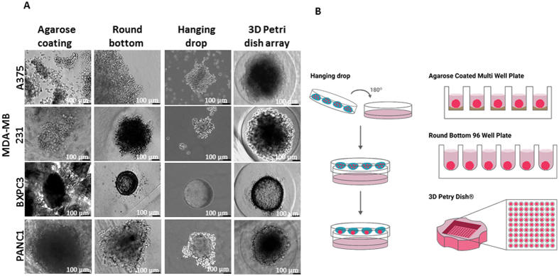
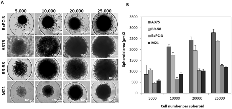
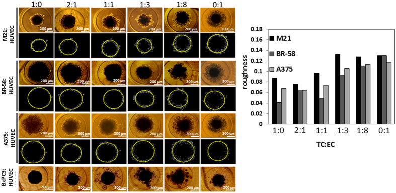
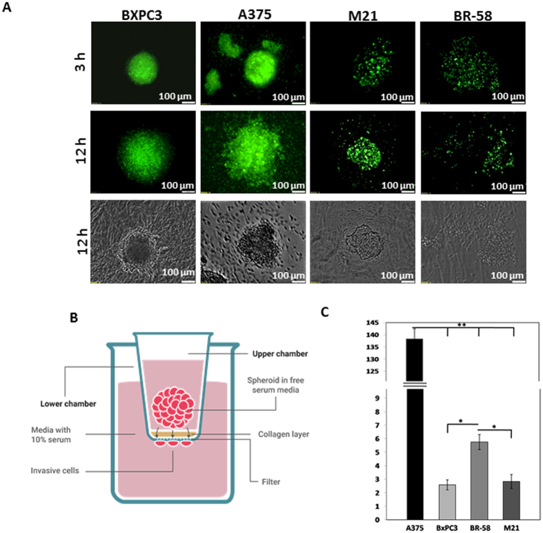
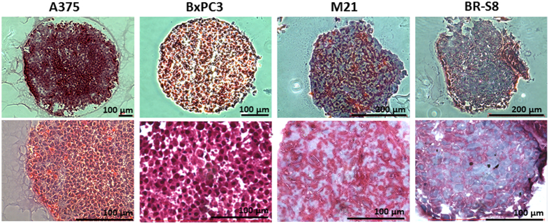
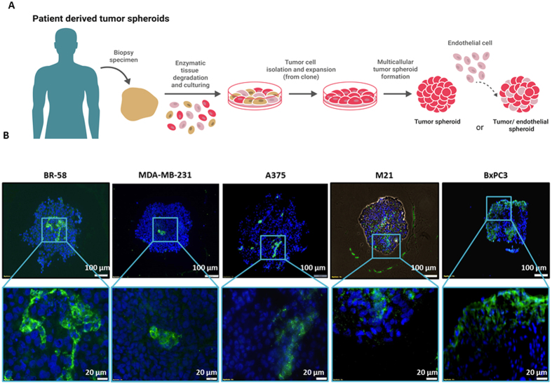
Recapitulating the tumor microenvironment is a central challenge in the development of experimental model for cancer. To provide a reliable tool for drug development and for personalized cancer therapy, it is critical to maintain key features that exist in the original tumor. Along with this effort, 3-dimentional (3D) cellular models are being extensively studied. Spheroids are self-assembled cell aggregates that possess many important components of the physiological spatial growth and cell-cell interactions. In this study we aimed to investigate the interconnection between tumor and endothelial cells (EC) in hybrid spheroids containing either tumor cell (TC) lines or patient derived cancer cells. Preparation protocols of hybrid spheroids were optimized and their morphology and tissue-like features were analyzed. Our finding show that capillary-like structures are formed upon assembly and growth of TC:EC spheroids and that spheroids' shape and surface texture may be an indication of spatial invasiveness of cells in the extra-cellular matrix (ECM). Establishing a model of hybrid tumor/stroma spheroids has a crucial importance in the experimental approach for personalized medicine, and may offer a reliable and low-cost method for the goal of predicting drug effects.


Conflict of interest statement

The authors declare that they have no competing interests.

Figures

Figure 1
Figure 1
Spheroids growth using different techniques, a comparison between spheroids that were prepared using the following formats: 96 well plate with agarose; Rounded bottom 96 well plate; 3D petri dish with 35 wells; Hanging drop method. Spheroids were made of 5,000 cells per spheroid. Assembly time was 3 days for all methods except the hanging drop method which was 5 days. Bar = 100 µm.
Figure 2
Figure 2
Spheroids made of patient tumor cells or tumor cell lines. (A) Typical images of spheroids made of BxPC3, A375, BR-58, and M21. Spheroids were fabricated using different number of cells per spheroid: 5,000, 10,000, 20,000 and 25,000. The spheroids were prepared using the 3D petri dish with 35 well array. (B) Graph showing spheroid area values (µm)2 as a function of the number of cells per spheroid. n = 4, Bar = 100 µm.
Figure 3
Figure 3
Tumor/Endothelial cell hybrid spheroids. Spheroids’ surface roughness (χ) values were calculated for spheroids composed of M21, BR-58 or A375 with different content of HUVEC cells. Typical images of hybrid spheroids and their corresponding MATLAB image analysis for roughness calculation. In the upper panels are the original images, and under each panel are the spheroid borders (white) with a fitting circle (yellow). BxPC3 spheroids were not analyzed due to satellite spheroid formation. Bar = 100 µm.
Figure 5
Figure 5
Spatial spheroid invasion assays: (A) 3D cultured spheroid made of BxPC3, A375, M21 and BR-58 embedded in collagen type I after 3 and 12 hours (bar = 100 µm). (B) Illustration of transwell invasion assay. In this assay, spheroids are seeded onto collagen coated polycarbonate membrane. The upper chamber contains serum free media and the lower chamber is supplemented with 10% serum. After 48 hour invasive cells that have detached form the spheroids are stained and counted. (C) Quantification of cell invasion from spheroids of BxPC3, A375, M21 and BR-58 in transwell assay. n = 12, **p < 0.025, *p < 0.05.
Figure 6
Figure 6
Histological staining for Collagen level in spheroid sections. Masson’s trichrome staining as performed in frozen sections of BR-58, M21, BxPC3 and A375 spheroids. Collagen is stained in blue. Bar = 100 µm.
Figure 4
Figure 4
Patient derived spheroids formation process and histological examination of TC:EC hybrid spheroids. (A) Illustration showing the isolation of tumor cells from a patient surgical sample, and assembly of tumor spheroids or TC:EC hybrid spheroids. (B)Immunofluorescent staining of EC using anti-CD31 in spheroid frozen sections of BR-58, M21, BxPC3 and A375 mixed with HUVEC at a 1:1 ratio. Green = CD31; Blue = nuclei DAPI staining.

References

    1. Ahn DH, Ciombor KK, Mikhail S, Bekaii-Saab T. Genomic diversity of colorectal cancer: Changing landscape and emerging targets. World journal of gastroenterology. 2016;22:5668–5677. doi: 10.3748/wjg.v22.i25.5668. - DOI - PMC - PubMed
    1. Niu B, et al. Protein-structure-guided discovery of functional mutations across 19 cancer types. Nature genetics. 2016;48:827–837. doi: 10.1038/ng.3586. - DOI - PMC - PubMed
    1. Kar SP, et al. Genome-Wide Meta-Analyses of Breast, Ovarian, and Prostate Cancer Association Studies Identify Multiple New Susceptibility Loci Shared by at Least Two Cancer Types. Cancer discovery. 2016 - PMC - PubMed
    1. Ishikawa T, et al. Emerging new technologies in Pharmacogenomics: rapid SNP detection, molecular dynamic simulation, and QSAR analysis methods to validate clinically important genetic variants of human ABC Transporter ABCB1 (P-gp/MDR1) Pharmacology & therapeutics. 2010;126:69–81. doi: 10.1016/j.pharmthera.2010.01.005. - DOI - PubMed
    1. Sotiriou C, Pusztai L. Gene-expression signatures in breast cancer. The New England journal of medicine. 2009;360:790–800. doi: 10.1056/NEJMra0801289. - DOI - PubMed

Publication types

LinkOut - more resources