. 2017 Sep 5;7(1):10541.
doi: 10.1038/s41598-017-10940-8.

Armored CAR T cells enhance antitumor efficacy and overcome the tumor microenvironment

Affiliations

Armored CAR T cells enhance antitumor efficacy and overcome the tumor microenvironment

Oladapo O Yeku et al. Sci Rep. .

Abstract

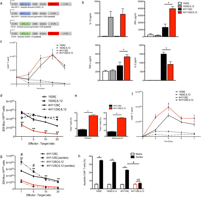
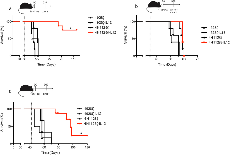
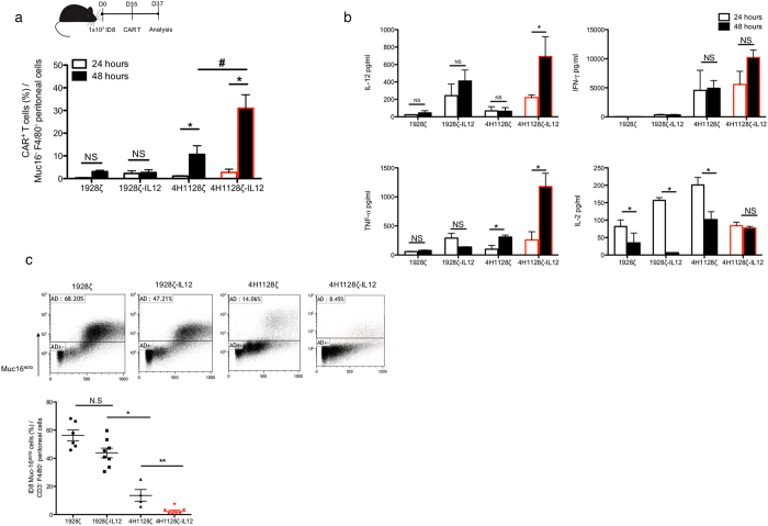
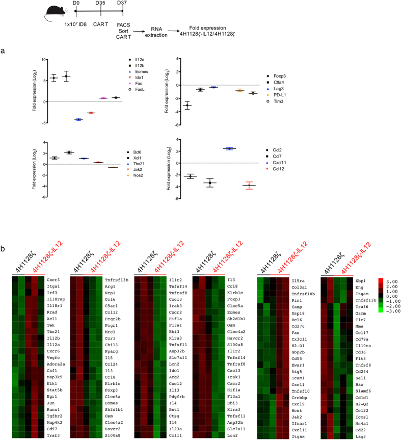
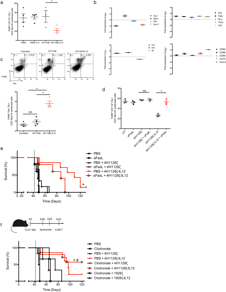
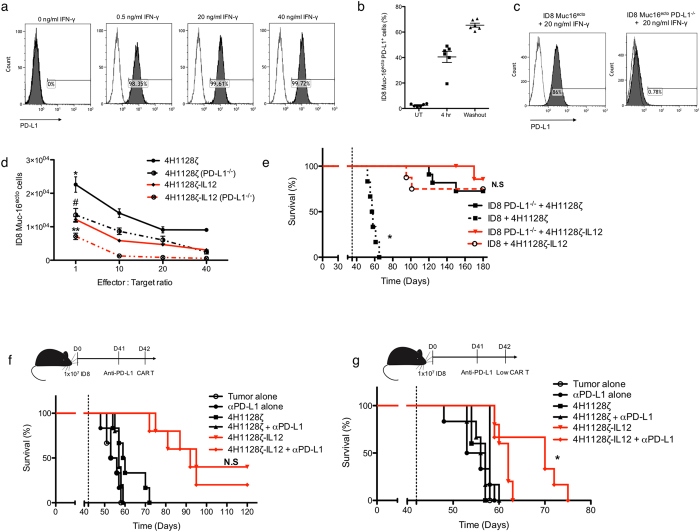
Chimeric antigen receptor (CAR) T cell therapy has shown limited efficacy for the management of solid tumor malignancies. In ovarian cancer, this is in part due to an immunosuppressive cytokine and cellular tumor microenvironment which suppresses adoptively transferred T cells. We engineered an armored CAR T cell capable of constitutive secretion of IL-12, and delineate the mechanisms via which these CAR T cells overcome a hostile tumor microenvironment. In this report, we demonstrate enhanced proliferation, decreased apoptosis and increased cytotoxicity in the presence of immunosuppressive ascites. In vivo, we show enhanced expansion and CAR T cell antitumor efficacy, culminating in improvement in survival in a syngeneic model of ovarian peritoneal carcinomatosis. Armored CAR T cells mediated depletion of tumor associated macrophages and resisted endogenous PD-L1-induced inhibition. These findings highlight the role of the inhibitory microenvironment and how CAR T cells can be further engineered to maintain efficacy.


Conflict of interest statement

Renier J. Brentjens is a co-founder, stockholder and consultant for Juno Therapeutics Inc.

Figures

Figure 1
Figure 1
Muc16ecto specific CAR T cells modified to secrete IL-12 elaborate more inflammatory cytokines, exhibit improved proliferation, increased cytotoxicity and decreased apoptosis in vitro. (a) Schematic representations of Muc16ecto- directed second generation CAR (4H1128ζ) modified to express murine IL-12 following an IRES element (4H1128ζ-IL12), and irrelevant CD19- directed second generation (1928ζ) and IL-12 modified (1928ζ-IL12) CARs. (b) In vitro cytokine analysis of supernatants obtained from coculture of indicated CAR T cells with ID8-Muc16ecto cells for 16 hr. IFN-γ: 4H1128ζ-IL12 vs 4H1128ζ, *p = 0.003. TNF-α: 4H1128ζ-IL12 vs 4H1128ζ CAR T cells, *p = 0.012. IL-2: 4H1128ζ-IL12 vs 4H1128ζ, *p = 0.045. Data are plotted as mean ± SEM (c). CAR T cell proliferation assay with indicated CAR T cells cocultured with ID8-Muc16ecto cells. (d) In vitro cytotoxicity assay of indicated CARs cocultured with ID8-Muc16ecto for 16 hr at the indicated effector: target ratios (E:T) on the x-axis, **p < 0.001. (e) Expression levels of perforin and granzyme B in 4H1128ζ-IL12 vs 4H1128ζ CAR T cells, *p < 0.0001 (f). CAR T cell proliferation assay with indicated CAR T cells cocultured with ID8-Muc16ecto cells in the presence of cell-free pooled ascites. 24 hr (*p < 0.001), 48 hr (*p = 0.046), 5 days (*p = 0.039). (g) In vitro cytotoxicity assay of indicated CARs cocultured with ID8-Muc16ecto for 16 hr in the presence of cell-free pooled ascites. 4H1128ζ-IL12 vs 4H1128ζ CAR T cells in ascites (*p < 0.01). 4H1128ζ vs 4H1128ζ ascites (#p < 0.01). (h) Indicated CAR T cells cocultured with ID8-Muc16ecto cells for 48 hr in the presence of complete media or ascites. Cells were gated on CAR T+ cells prior to gating on annexin V/DAPI. *p < 0.01, #p < 0.01. Data are plotted as mean ± SEM. Data shown are pooled results from 3 independent experiments. Statistics performed using unpaired two-sided T test.
Figure 2
Figure 2
IL-12-secreting CAR T cells prolong survival in an advanced tumor model of ovarian peritoneal carcinomatosis and is dependent on autocrine IL-12 signaling. (a) Female wild type C57BL/6 mice (WT) between 6–8 weeks old were inoculated with 1 × 107 ID8-Muc16ecto cells i.p and treated 35 days later with 2 × 106 indicated CAR T cells i.p., *p = 0.02. Number of mice per group: 5 (1928ζ), 5 (1928ζ-IL12), 6 (4H1128ζ), 8 (4H1128ζ-IL12). (b) Female WT mice between 6–8 weeks old were inoculated with 1 × 107 ID8-Muc16ecto cells i.p and treated 35 days later with 2 × 106 CAR T cells (i.p) derived from IL-12R−/− splenocytes. Number of mice per group: 5 (1928ζ), 5 (1928ζ-IL12), 5 (4H1128ζ), 5 (4H1128ζ-IL12). (c) Female WT mice between 6–8 weeks old were inoculated with 1 × 107 ID8-Muc16ecto cells i.p and treated 42 days later with 2 × 106 indicated CAR T cells i.p, *p < 0.01. Number of mice per group: 5 (1928ζ), 5 (1928ζ-IL12), 6 (4H1128ζ), 8 (4H1128ζ-IL12). Results are pooled from 2 independent experiments. Statistical analysis performed using a log-rank (Mantel-Cox) test.
Figure 3
Figure 3
4H1128ζ-IL12 CAR T cells expand better, secrete inflammatory cytokines and eradicate tumor cells in vivo. (a) Female WT mice between 6–8 weeks old were inoculated with 1 × 107 ID8-Muc16ecto cells i.p and treated 35 days later with 2 × 106 CAR T cells i.p. Mice were subsequently euthanized 24 hr and 48 hr after CAR T cell injection and peritoneal washes were performed. FACS analysis was performed on recovered peritoneal cells and CAR+ T cells were gated on Muc16 and F4/80 cells. 4H1128ζ (*p = 0.01), 4H1128ζ-IL12 (*p = 0.04), #p < 0.05. Data are plotted as mean ± SEM. Data shown are pooled results from 3 independent experiments. (b) Cytokine analysis performed on peritoneal fluid samples obtained from D35 tumor-bearing mice treated with CAR T cells as in (a). IL-12: (*p < 0.01), TNF-α (*p < 0.01), and IL-2: 1928ζ (*p = 0.031), 1928ζ-IL12 (*p < 0.01), 4H1128ζ (*p < 0.01). Data are plotted as mean ± SEM. Results are pooled from 3 independent experiments. (c) WT female mice between 6–8 weeks old inoculated with 1 × 107 ID8-Muc16ecto cells i.p were treated 35 days later with 2 × 106 CAR T cells i.p and subjected to peritoneal fluid analysis 48 hr after T cell infusion for the number of viable ID8-Muc16ecto cells. *p < 0.05, **p < 0.001. Data are plotted as mean ± SEM. Results are pooled from 3 independent experiments. Statistics performed using unpaired two-sided T test.
Figure 4
Figure 4
4H1128ζ-IL12 CAR T cells express decreased exhaustion markers. (a) Differential expression of statistically significant (p < 0.05) genes between pooled recovered CAR T cells from 4H1128ζ-IL12 and 4H1128ζ- treated mice. mRNA was extracted from pooled CAR T cells from 2 mice and performed in duplicate for 4H1128ζ and in triplicate for 4H1128ζ-IL12. n = 4 (4H1128ζ), n = 6 (4H1128ζ-IL12). Data presented as fold change of 4H1128ζ-IL12/4H1128ζ and plotted as mean ± SEM. (b) Heatmap representation of differentially expressed genes between recovered 4H1128ζ-IL12 and 4H1128ζ CAR T cells.
Figure 5
Figure 5
4H1128ζ-IL12 CAR T cells phenotypically alter and delete tumor associated macrophages. (a) Peritoneal washes from tumor-bearing mice treated with 4H1128ζ-IL12 compared to 4H1128ζ CAR T cells and assessed for F4/80+ CD11b+ TAM (*p < 0.05). Data are plotted as mean ± SEM. Data shown are pooled results from 3 independent experiments. (b) Statistically significant (p < 0.05) differential gene expression between F4/80+ CD11b+ TAM recovered from mice treated with 4H1128ζ-IL12 vs 4H1128ζ. mRNA was extracted from pooled TAM from 2 mice per group after FACS sorting. n = 6 (4H1128ζ), n = 6 (4H1128ζ-IL12). Data presented as fold change of 4H1128ζ-IL12 TAM / 4H1128ζ TAM and plotted as mean ± SEM. (c) Analysis of Fas expression on F4/80+ TAM recovered from animals treated with CAR T cells (*p < 0.01). Data are plotted as mean ± SEM. Data shown are pooled results from 3 independent experiments. (d) TAM recovery from day 35 tumor-bearing mice treated with anti-FasL (αFasL) neutralizing antibody 1 day prior to CAR T infusion, *p < 0.01. Data are plotted as mean ± SEM. Data shown are pooled results from 3 independent experiments. (e) Female WT mice age 6–8 weeks inoculated with 1 × 107 ID8-Muc16ecto cells i.p followed by treatment with 250 μg αFasL i.p on day 41 and CAR T cells on day 42. *p = 0.04. Number of mice per group: 5 (PBS), 5 (αFasL), 6 (PBS + 4H1128ζ), 6 (αFasL + 4H1128ζ), 7 (PBS + 4H1128ζ-IL12), 6 (αFasL + 4H1128ζ-IL12). Results are pooled from 2 independent experiments. (f) WT tumor-bearing female mice between 6–8 weeks old were treated with 2 doses of clodronate (Clod) or PBS liposomes on Day 38 and 40. Subsequently, mice were treated with CAR T cells on D42. *p < 0.05. Number of mice per group: 5 (PBS), 5 (Clod), 5 (PBS + 4H1128ζ), 5 (PBS + 4H1128ζ-IL12), 6 (Clod + 4H1128ζ), 7 (Clod + 4H1128ζ-IL12), 5 (Clod + 1928ζ), 5 (Clod + 1928ζ-IL12). Results are pooled from 3 independent experiments. Statistical analysis performed using a log-rank (Mantel-Cox) test.
Figure 6
Figure 6
4H1128ζ-IL12 CAR T cells overcome endogenous tumor PD-L1 suppression. (a) ID8-Muc16ecto cells cocultured with various concentrations of recombinant IFN-γ and incubated for 16 hr. (b) ID8-Muc16ecto cells incubated with recombinant IFN-γ and assessed for PD-L1 upregulation after 4 hr of exposure. Similarly, tumor cells were exposed to IFN-γ for 16 hr, washed and then incubated in media without IFN-γ for 24 hr. Data are plotted as mean ± SEM. Data shown are pooled from 2 independent experiments. (c) Murine PD-L1 gene was genetically deleted using CRISPR-Cas9 and ID8-Muc16ecto PD-L1−/− cells were screened via stimulation with IFN-γ for 16 hr. Data shown are pooled from 2 independent experiments. (d) ID8-Muc16ecto PD-L1−/− and ID8-Muc16ecto cells cocultured with CAR T cells at the indicated E:T ratios for 16 hr. The number of residual tumor cells from each condition is shown, *p < 0.05, **p < 0.05, #N.S. Data shown are pooled from 2 independent experiments. (e) D42 tumor-bearing mice inoculated with ID8-Muc16ecto or ID8-Muc16ecto PD-L1−/− cells and treated with indicated CAR T cells. *p < 0.001. n = 8 mice per group. Results pooled from 2 independent experiments. (f) D42 tumor-bearing mice previously inoculated with ID8-Muc16ecto cells were pretreated on D41 with 250 μg of anti-PD-L1 blocking antibody (αPD-L1) prior to i.p infusion of 2 × 106 4H1128ζ-IL12 or 4H1128ζ CAR T cells. Number of mice per group: 3 (Tumor), 6 (αPD-L1), 6 (4H1128ζ), 6 (4H1128ζ + αPD-L1), 6 (4H1128ζ-IL12), 6 (4H1128ζ-IL12 + αPD-L1). (g) D42 tumor-bearing mice pretreated with 250 μg αPD-L1 followed by 2 × 105 CAR T cells, *p < 0.05. Number of mice per group: 3 (Tumor), 6 (αPD-L1), 5 (4H1128ζ), 6 (4H1128ζ + αPD-L1), 5 (4H1128ζ-IL12), 6 (4H1128ζ-IL12 + αPD-L1). Results are pooled from 2 independent experiments. Statistical analysis performed using a log-rank (Mantel-Cox) test.

References

    1. Davila ML, et al. Chimeric antigen receptors for the adoptive T cell therapy of hematologic malignancies. Int. J. Hematol. 2014;99:361–71. doi: 10.1007/s12185-013-1479-5. - DOI - PMC - PubMed
    1. Park JR, et al. Adoptive transfer of chimeric antigen receptor re-directed cytolytic T lymphocyte clones in patients with neuroblastoma. Mol. Ther. 2007;15:825–33. doi: 10.1038/sj.mt.6300104. - DOI - PubMed
    1. Feng K, et al. Chimeric antigen receptor-modified T cells for the immunotherapy of patients with EGFR-expressing advanced relapsed/refractory non-small cell lung cancer. Sci. China. Life Sci. 2016;59:468–79. doi: 10.1007/s11427-016-5023-8. - DOI - PubMed
    1. Beatty GL, et al. Mesothelin-specific chimeric antigen receptor mRNA-engineered T cells induce anti-tumor activity in solid malignancies. Cancer Immunol. Res. 2014;2:112–20. doi: 10.1158/2326-6066.CIR-13-0170. - DOI - PMC - PubMed
    1. Kershaw MH, et al. A phase I study on adoptive immunotherapy using gene-modified T cells for ovarian cancer. Clin. Cancer Res. 2006;12:6106–15. doi: 10.1158/1078-0432.CCR-06-1183. - DOI - PMC - PubMed

Publication types