. 2017 Sep 5;7(1):10446.
doi: 10.1038/s41598-017-11036-z.

SRSF1 suppresses selection of intron-distal 5' splice site of DOK7 intron 4 to generate functional full-length Dok-7 protein

Affiliations

SRSF1 suppresses selection of intron-distal 5' splice site of DOK7 intron 4 to generate functional full-length Dok-7 protein

Khalid Bin Ahsan et al. Sci Rep. .

Abstract

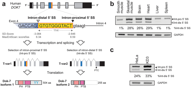
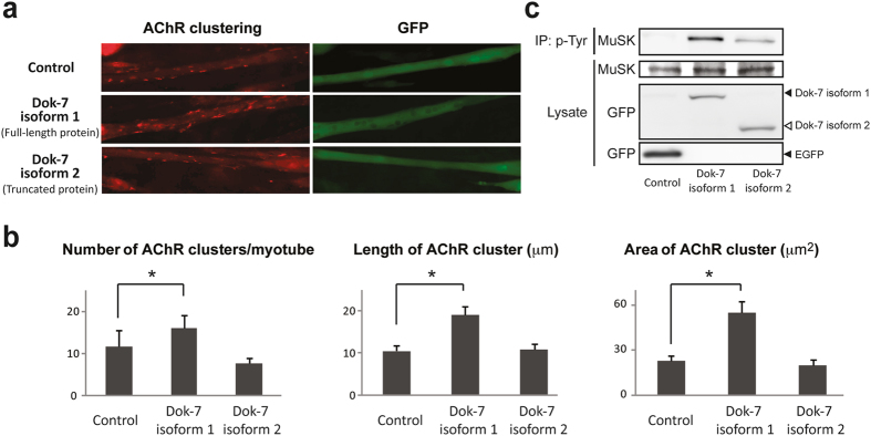
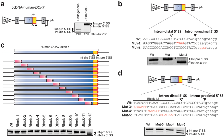
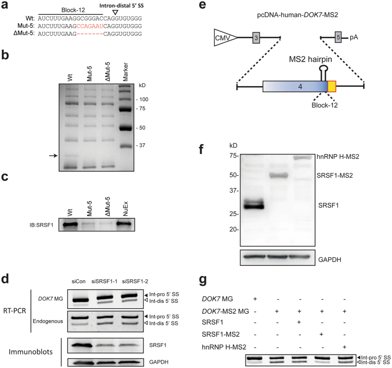
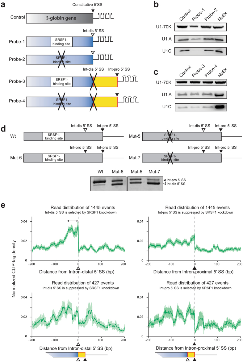
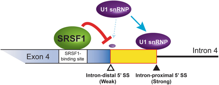
Dok-7 is a non-catalytic adaptor protein that facilitates agrin-induced clustering of acetylcholine receptors (AChR) at the neuromuscular junction. Alternative selection of 5' splice sites (SSs) of DOK7 intron 4 generates canonical and frame-shifted transcripts. We found that the canonical full-length Dok-7 enhanced AChR clustering, whereas the truncated Dok-7 did not. We identified a splicing cis-element close to the 3' end of exon 4 by block-scanning mutagenesis. RNA affinity purification and mass spectrometry revealed that SRSF1 binds to the cis-element. Knocking down of SRSF1 enhanced selection of the intron-distal 5' SS of DOK7 intron 4, whereas MS2-mediated artificial tethering of SRSF1 to the identified cis-element suppressed it. Isolation of an early spliceosomal complex revealed that SRSF1 inhibited association of U1 snRNP to the intron-distal 5' SS, and rather enhanced association of U1 snRNP to the intron-proximal 5' SS, which led to upregulation of the canonical DOK7 transcript. Integrated global analysis of CLIP-seq and RNA-seq also indicated that binding of SRSF1 immediately upstream to two competing 5' SSs suppresses selection of the intron-distal 5' SS in hundreds of human genes. We demonstrate that SRSF1 critically regulates alternative selection of adjacently placed 5' SSs by modulating binding of U1 snRNP.


Conflict of interest statement

The authors declare that they have no competing interests.

Figures

Figure 1
Figure 1
Schematic of alternative splicing of DOK7, and its splicing pattern in human tissues and cell lines. (a) Schematic of genomic structure of human DOK7 gene locus. Exons and introns are shown in boxes and solid lines, respectively. Human DOK7 intron 4 contains two 5′ SSs, which are 11 nucleotides apart. Open and closed arrowheads point to the intron-distal and intron-proximal 5′ SSs, respectively. Constitutive segment of exon 4 is indicated by a blue box, and alternatively spliced segment of exon 4 is indicated by a yellow box. Transcript variant 1 (T-var1) and transcript variant 2 (T-var2) are schematically shown below the gene structure. Position of the premature terminal codon (PTC) in T-var2 is indicated by an arrow. T-var1 encodes the canonical full-length Dok-7 isoform 1 containing the PH and PTB domains, as well as two tyrosine residues (YY) that are target motifs of SH2 domain. T-var2 encodes the truncated Dok-7 isoform 2 retaining the PH domain and part of the PTB domain, but lacking two tyrosine residues. (b) RT-PCR showing alternative 5′ SS selection of DOK7 intron 4 in human tissues. DOK7 transcripts are highly expressed in the skeletal muscle, brain, and heart, in which both the intron-proximal 5′ SS (Int-pro 5′ SS) and the intron-distal 5′ SS (Int-dis 5′ SS) are selected. (c) RT-PCR showing alternative 5′ SS selection of DOK7 intron 4 in HeLa cells and immortalized human myogenic KD3 cells.
Figure 2
Figure 2
Functional assay of Dok-7 isoforms 1 and 2 in AChR clustering. (a) Enhancement of agrin-mediated AChR clustering of C2C12 myotubes by overexpression of Dok-7 isoforms 1 and 2. C2C12 myoblasts are transfected with cDNA encoding Dok-7 isoform 1 or 2 fused with GFP, or cDNA encoding GFP alone (Control), and are cultured in differentiation medium. On day 5, 10 ng/ml agrin is added to induce AChR clustering, and AChR clustering is visualized with Alexa594-conjugated α-bungarotoxin at 12 h later. Transfected C2C12 myotubes are identified by GFP. (b) Morphometric analysis showing that Dok-7 isoform 1, but not isoform 2, induces AChR clustering. The mean and S.E. are indicated. p < 0.05 (not indicated) by one-way ANOVA for all datasets. *p < 0.05 by post-hoc Tukey HSD test. (c) MuSK phosphorylation assay of differentiation-induced C2C12 myoblasts transfected with cDNA encoding Dok-7 isoform 1 or 2 fused with GFP, or cDNA encoding GFP alone (Control), along with cDNA encoding FLAG-tagged MuSK. Phosphorylated MuSK is detected by immunoprecipitation of cell lysate by anti-phosphotyrosine antibody (p-Tyr) followed by immunoblotting with anti-FLAG antibody (MuSK).
Figure 3
Figure 3
Identification of a splicing regulatory cis-element in DOK7 exon 4. (a) Structure of human DOK7 minigene spanning exons 3 to 5 driven by a CMV promoter (pcDNA-human-DOK7). Positions of intron-distal (Int-dis) and intron-proximal (Int-pro) 5′ SS are indicated by open and closed arrowheads, respectively. Positions of primers for RT-PCR are indicated by bent arrows. RT-PCR of endogenous DOK7 transcripts and DOK7 minigene transcripts (DOK7 MG) in HeLa cells are shown in the right panel. Percentages of the selection of intron-distal 5′ SS are shown at the bottom. (b) Schematic of pcDNA-human-DOK7 minigenes carrying wild-type (Wt) and mutant (Mut-1 and -2) 5′ SSs. Introduced mutations are shown in red. RT-PCR of wild-type and mutant constructs transfected in HeLa cells are shown. A transcript generated by activation of a cryptic splice site at IVS4 + 12 (not shown) in intron 4 is indicated by an asterisk. (c) Schematic of cis-regulatory block-scanning mutagenesis of DOK7 exon 4 in the context of pcDNA-human-DOK7 minigene. A 15-nucleotide heterologous sequence of 5′-TCAGTATGACTCTCA-3′ is introduced into each block, excluding Block-12, which is replaced with 19-nucleotide heterologous sequence of 5′-TCAGTATGACTCTCAGTAT-3′. No mutations are introduced into the first three nucleotides and the last fourteen nucleotides of exon 4, where the intron-distal 5′ SS is located. Positions of intron-distal (Int-dis) and intron-proximal (Int-pro) 5′ SS are indicated by open and closed arrowheads, respectively. RT-PCR of these constructs in HeLa cells are shown below. (d) Schematic of pcDNA-human-DOK7 minigenes carrying wild-type (Wt) and mutant (Mut-3, -4, and -5) sequence. RT-PCR of wild-type and mutant constructs transfected in HeLa cells are shown. (b,d) Mutant nucleotides are indicated in red. Uppercase and lowercase letters represent exonic and intronic nucleotides, respectively.
Figure 4
Figure 4
SRSF1 binds to the identified cis-element and facilitates selection of the intron-proximal 5′ SS of DOK7 intron 4. (a) RNA probes carrying wild-type (Wt), mutant (Mut-5), and partially deleted (ΔMut-5) sequences. (b) Coomassie blue staining of RNA affinity-purified products using HeLa nuclear extract with the indicated biotinylated RNA probes. A single protein band at ~30 kDa (black arrow) is associated with Wt probe, but not with Mut-5 or ΔMut-5 probe. Mass spectrometry analysis revealed that the identity of this protein is SRSF1. The other bands commonly observed in the Wt, Mut-5 and ΔMut-5 RNA probes are repeatedly identified in our RNA affinity purification analyses, , . Mass spectrometry analysis of these bands revealed that none of the identified proteins carry an RNA-recognition motif, suggesting that these bands are likely due to non-specific binding of proteins to the streptavidin-sepharose beads. NuEx, HeLa nuclear extract. (c) Immunoblotting of RNA affinity-purified proteins in panel (b) with anti-SRSF1 antibody. (d) Upper panels show RT-PCR of DOK7 minigene (DOK7 MG) encoded by pcDNA-human-DOK7 and endogenous DOK7 transcripts in HeLa cells treated with two different siRNAs against SRSF1 (siSRSF1-1 and siSRSF1-2). Lower panels show immunoblotting with indicated antibodies after SRSF1-knockdown with two different siRNAs in HeLa cells. (e) Schematic of pcDNA-human-DOK7-MS2 minigene. Block-12 sequence is substituted with the MS2 coat protein-binding hairpin RNA sequence. (f) Immunoblotting with anti-His antibody to show overexpression of His-tagged transgene products in HeLa cells: SRSF1, SRSF1 fused with MS2 coat protein (SRSF1-MS2), and hnRNP H fused with MS2 coat protein (hnRNP H-MS2). (g) RT-PCR of DOK7 minigene (DOK7 MG) and DOK7-MS2 minigene (DOK7-MS2 MG) encoded by pcDNA-human-DOK7 and pcDNA-human-DOK7-MS2, respectively, in HeLa cells co-transfected with the indicated effectors. Int-pro 5′ SS and Int-dis 5′ SS point to transcripts generated by selection of the intron-proximal and intron-distal 5′ SSs, respectively.
Figure 5
Figure 5
SRSF1 suppresses binding of U1 snRNP to the intron-distal 5′ SS of DOK7 intron 4. (a) Schematic of 3 x MS2 RNA probes used for isolation of an early spliceosome complex. Control probe contains constitutive 5′ SS of the human β-globin gene encoded in pSP64-HβΔ6-MS2. Probe-1 carries the SRSF1-binding site and the intron-distal (Int-dis) 5′ SS, but lacks the intron-proximal (Int-pro) 5′ SS. Probe-2 carries a disrupted SRSF1-binding site and the intron-distal 5′ SS, but lacks the intron-proximal 5′ SS. Probe-3 carries the SRSF1-binding site, a mutated intron-distal 5′ SS, and the intron-proximal 5′ SS. Probe-4 carries a disrupted SRSF1-binding site, a mutated intron-distal 5′ SS, and the intron-proximal 5′ SS. Sequences of the probes are shown in Supplementary Fig. S3. (b) Immunoblotting of purified spliceosome complexes assembled on control, probe-1, and probe-2 RNA substrates. (c) Immunoblotting of purified spliceosome complexes assembled on control, probe-3, and probe-4 RNA substrates. (d) Schematic of DOK7 minigenes carrying wild-type (Wt and Mut-6) or disrupted (Mut-5 and Mut-7) SRSF1-binding site, with (Mut-6 and Mut-7) or without (Wt and Mut-5) duplicated intron-proximal 5′ SS. Lower panel shows RT-PCR of these minigenes transfected in HeLa cells. Sequences of the Wt and mutant constructs (Mut-5, -6 and -7) are shown in Supplementary Fig. S6. Mut-5 is identical to Mut-5 in Fig. 3d. (e) Distribution of SRSF1-CLIP tags centered around the intron-distal (left panels) and intron-proximal (right panels) 5′ SSs depicted by integrated genome-wide analysis of CLIP-seq of SRSF1 in native HeLa cells and RNA-seq in SRSF1-knocked down HeLa cells. Mean (green lines) and standard error (light green areas) of normalized CLIP-tag densities are shown. In 1445 (upper panels) and 427 (lower panels) genes, SRSF1-konockdown activates intron-distal and intron-proximal 5′ SSs, respectively. A double-headed arrow indicates a peak immediately upstream to the intron-distal 5′ SS in the 1445 genes, indicating the suppressive effect of SRSF1 on the intron-distal 5′ SS, as we observed in DOK7 intron 4.
Figure 6
Figure 6
Schematic of alternative 5′ SS selection of human DOK7 intron 4. Binding of SRSF1 immediately upstream to the intron-distal 5′ SS suppresses binding of U1 snRNP to the intron-distal 5′ SS, but not to the intron-proximal 5′ SS.

Similar articles

Cited by

References

    1. Pan Q, Shai O, Lee LJ, Frey BJ, Blencowe BJ. Deep surveying of alternative splicing complexity in the human transcriptome by high-throughput sequencing. Nat Genet. 2008;40:1413–1415. doi: 10.1038/ng.259. - DOI - PubMed
    1. Ohno, K. et al. Splicing regulation and dysregulation of cholinergic genes expressed at the neuromuscular junction. Journal of Neurochemistry (2017). - PubMed
    1. Nakai K, Sakamoto H. Construction of a novel database containing aberrant splicing mutations of mammalian genes. Gene. 1994;141:171–177. doi: 10.1016/0378-1119(94)90567-3. - DOI - PubMed
    1. Brogna S, Wen J. Nonsense-mediated mRNA decay (NMD) mechanisms. Nat Struct Mol Biol. 2009;16:107–113. doi: 10.1038/nsmb.1550. - DOI - PubMed
    1. Cartegni L, Chew SL, Krainer AR. Listening to silence and understanding nonsense: exonic mutations that affect splicing. Nat Rev Genet. 2002;3:285–298. doi: 10.1038/nrg775. - DOI - PubMed

Publication types

LinkOut - more resources