. 2017 Sep 5;7(1):10522.
doi: 10.1038/s41598-017-11274-1.

Structural determinants of 5',6'-epoxyeicosatrienoic acid binding to and activation of TRPV4 channel

Affiliations

Structural determinants of 5',6'-epoxyeicosatrienoic acid binding to and activation of TRPV4 channel

Alejandro Berna-Erro et al. Sci Rep. .

Abstract

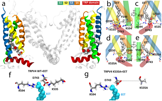
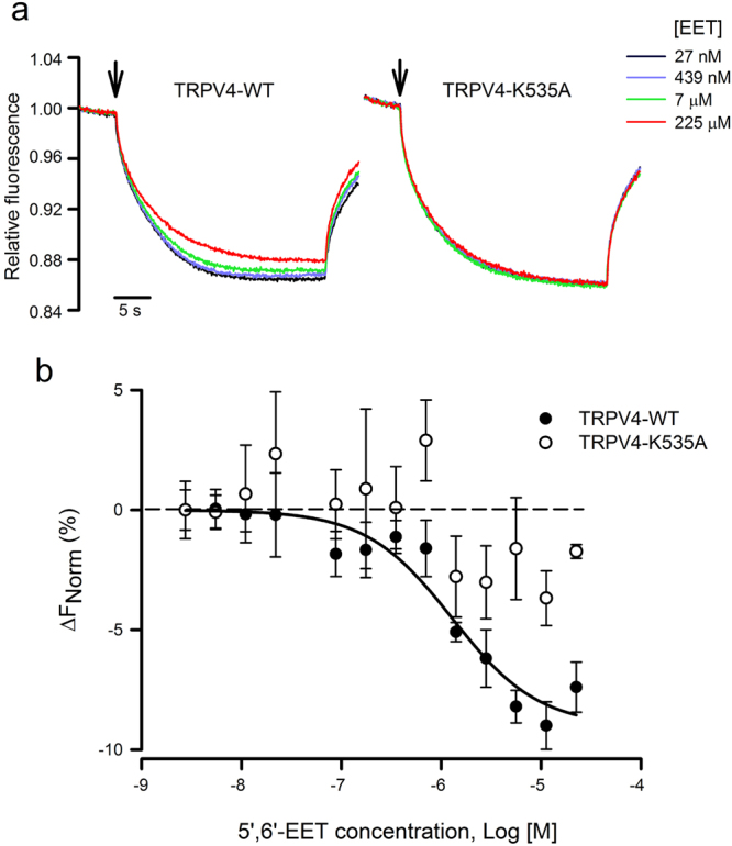
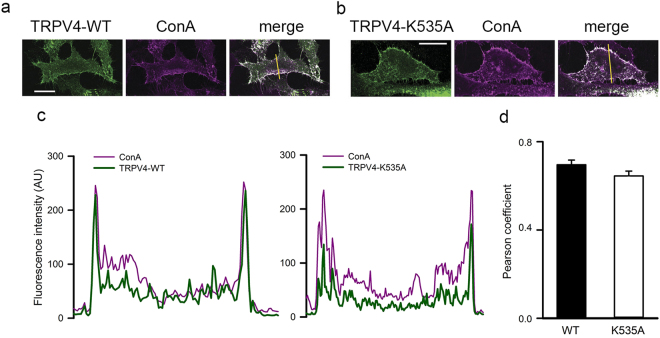
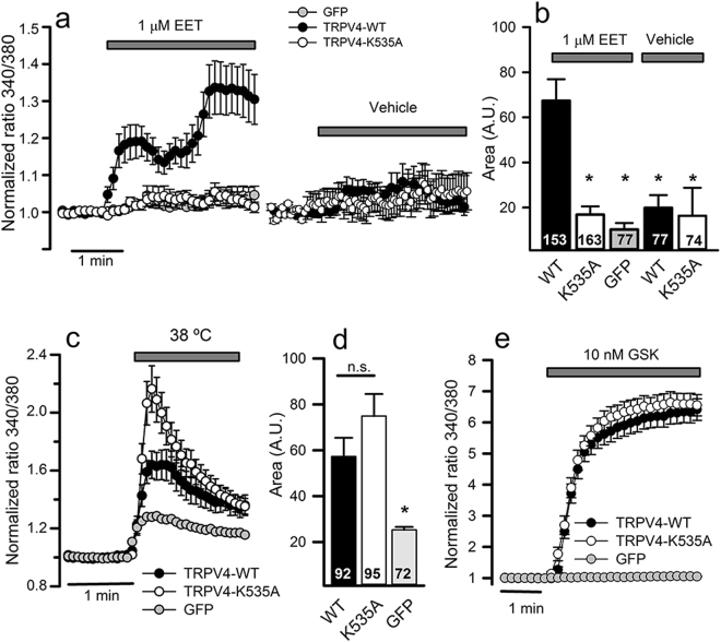
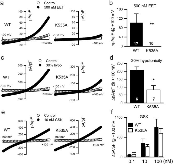
TRPV4 cation channel activation by cytochrome P450-mediated derivatives of arachidonic acid (AA), epoxyeicosatrienoic acids (EETs), constitute a major mechanisms of endothelium-derived vasodilatation. Besides, TRPV4 mechano/osmosensitivity depends on phospholipase A2 (PLA2) activation and subsequent production of AA and EETs. However, the lack of evidence for a direct interaction of EETs with TRPV4 together with claims of EET-independent mechanical activation of TRPV4 has cast doubts on the validity of this mechanism. We now report: 1) The identification of an EET-binding pocket that specifically mediates TRPV4 activation by 5',6'-EET, AA and hypotonic cell swelling, thereby suggesting that all these stimuli shared a common structural target within the TRPV4 channel; and 2) A structural insight into the gating of TRPV4 by a natural agonist (5',6'-EET) in which K535 plays a crucial role, as mutant TRPV4-K535A losses binding of and gating by EET, without affecting GSK1016790A, 4α-phorbol 12,13-didecanoate and heat mediated channel activation. Together, our data demonstrates that the mechano- and osmotransducing messenger EET gates TRPV4 by a direct action on a site formed by residues from the S2-S3 linker, S4 and S4-S5 linker.


Conflict of interest statement

The authors declare that they have no competing interests.

Figures

Figure 1
Figure 1
Predicted model of TRPV4 channel interaction with 5′,6′-EET. (a) TRPV4 structural model showing only two of the four identical subunits (side view). Color-coded transmembrane segments S1-S4, TRP box and EET molecule are highlighted. Images of the predicted EET-binding site in TRPV4-WT (b,c) and TRPV4 K535A (d,e) systems. Residues showing more than 60% of time occupancy are displayed in licorice representation. K535A residue from TRPV4 K535A is displayed in licorice representation even though its interaction is not significant. (f,g) Bottom view detail of the interaction of 5′,6′-EET with residues K535 and R594 in the TRPV4-WT (f) and TRPV4-K535A (g) systems. Note the loss of interaction between mutated residue A535 and both D743 and EET molecule.
Figure 2
Figure 2
Thermophoretic analysis of the TRPV4-EET interaction. (a) Typical signal of a microscale thermophoresis (MST) experiment showing the thermophoretic movement (induced by infrared laser activation, arrow) of the GFP-labeled TRPV4 WT or K535A mutant channel and the subsequent fluorescence change measured for 30 s in the presence of increasing concentrations of 5′,6′-EET, from 27 nM (blue trace) to 225 µM (red trace). GFP-TRPV4 channels and 5′,6′-EET were incubated for 30–60 min in PBS, with 2% ethanol as vehicle. (b) Average changes in normalized fluorescence obtained for GFP-labeled TRPV4 WT (n = 4) and K535 (n = 4) channels plotted against 5′,6′-EET concentration and fitting of the TRPV4-WT data.
Figure 3
Figure 3
Mutation K535A does not alter membrane localization of heterologously expressed TRPV4 channels. Co-localization of TRPV4-WT (a) and TRPV4-K535A (b) channels (green) expressed in HeLa cells with the plasma membrane marker concanavalin A (magenta). Co-localized pixels are shown in white in the merge panels. The yellow line on the merge panels indicates the plot profile analysis (c) performed on each image using ImageJ software. Scale bar = 20 μm. (d) Pearson correlation coefficients obtained with the plot profile analysis of TRPV4-WT and TRPV4-K535A channels in the plasma membrane location. Mean ± S.E.M., P = 0.1 Mann-Whitney.
Figure 4
Figure 4
TRPV4-K535A channel loses activation by 5′,6′-EET not by heat or GSK1016790A agonist. (a) Changes in intracellular [Ca2+] (indicated by normalized fura-2 ratios) in HeLa cells transfected with GFP, TRPV4-WT or TRPV4-K535A cDNAs, after perfusion with 1 µM 5′,6′-EET or vehicle (as indicated). Traces are means ± SEM of 74–163 cells measured in 4–8 independent experiments. (b) Average [Ca2+]i increases (area under the curve) from traces shown in a. Numbers inside the bars indicate the number of cells analyzed. (c) Changes in intracellular [Ca2+] in response to warm solution (38 °C). (d) Average [Ca2+]i increases (area under the curve) from traces shown in c. (e) Changes in intracellular [Ca2+] in response to 10 nM GSK1016790A (GSK). Traces are means of 93–109 cells measured in 4–6 independent experiments. *P < 0.05 or not significant (n.s.) when compared with cells expressing TRPV4 WT channels (Kruskal-Wallis followed by Dunn post hoc test).
Figure 5
Figure 5
Effect of K535A substitution on the activation of TRPV4 whole-cell currents. Representative ramp current desnsity-voltage relations of whole-cell cationic currents recorded from HEK293 cells transfected with either TRPV4 WT or TRPV4 K535A cDNAs before (control) and after dialysis with 500 nM 5′,6′-EET (a), exposition to a 30% hypotonic shock (c) or to 10 nM GSK1016790A (e). (b,d,f) Average TRPV4 current density (at + 100 mV) increases in response to the above indicated stimuli. Data are expressed as the mean ± SEM, and the number of cells recorded is shown for each experimental condition. **P < 0.01, *P < 0.05 K535A versus WT. P values in panel b = 0.007, panel d = 0.002 and > 0.05 at al concentrations in panel f (One-tailed Student′s unpaired t test).
Figure 6
Figure 6
K535A substitution prevents the 5′,6′-EET-induced change in TRPV4 pore dimension. Pore area delimited by the square formed by the four Cα of I715 residues (located at the narrowest zone of TRPV4 pore). (a) TRPV4-WT; (b) TRPV4-WT + 5′,6′-EET; (c) TRPV4-K535A; (d) TRPV4-K535A + 5′,6′-EET.
Figure 7
Figure 7
K535A substitution abolishes the 5′,6′-EET-induced water flux through TRPV4 pore. Water presence around I715 residue in TRPV4-WT (a), TRPV4-WT + EET (b), TRPV4-K535A (c), and TRPV4-K535A + EET (d) systems. Insets: Water molecules crossing the hydrophobic seal formed by I715 are observed exclusively in the TRPV4-WT + EET system.

References

    1. Garcia-Elias A, et al. The TRPV4 channel. Handb. Exp. Pharmacol. 2014;222:293–319. doi: 10.1007/978-3-642-54215-2_12. - DOI - PubMed
    1. White JPM, et al. TRPV4: Molecular Conductor of a Diverse Orchestra. Physiol. Rev. 2016;96:911–973. doi: 10.1152/physrev.00016.2015. - DOI - PubMed
    1. Strotmann R, Harteneck C, Nunnenmacher K, Schultz G, Plant TD. OTRPC4, a nonselective cation channel that confers sensitivity to extracellular osmolarity. Nat. Cell Biol. 2000;2:695–702. doi: 10.1038/35036318. - DOI - PubMed
    1. Liedtke W, et al. Vanilloid receptor-related osmotically activated channel (VR-OAC), a candidate vertebrate osmoreceptor. Cell. 2000;103:525–535. doi: 10.1016/S0092-8674(00)00143-4. - DOI - PMC - PubMed
    1. Arniges M, et al. Swelling-activated Ca2+ entry via TRPV4 channel is defective in cystic fibrosis airway epithelia. J. Biol. Chem. 2004;279:54062–54068. doi: 10.1074/jbc.M409708200. - DOI - PubMed

Publication types

Substances