. 2017 Sep 5;7(1):10542.
doi: 10.1038/s41598-017-10836-7.

Hyaluronic Acid Coated Chitosan Nanoparticles Reduced the Immunogenicity of the Formed Protein Corona

Affiliations

Hyaluronic Acid Coated Chitosan Nanoparticles Reduced the Immunogenicity of the Formed Protein Corona

Abdulaziz Almalik et al. Sci Rep. .

Abstract

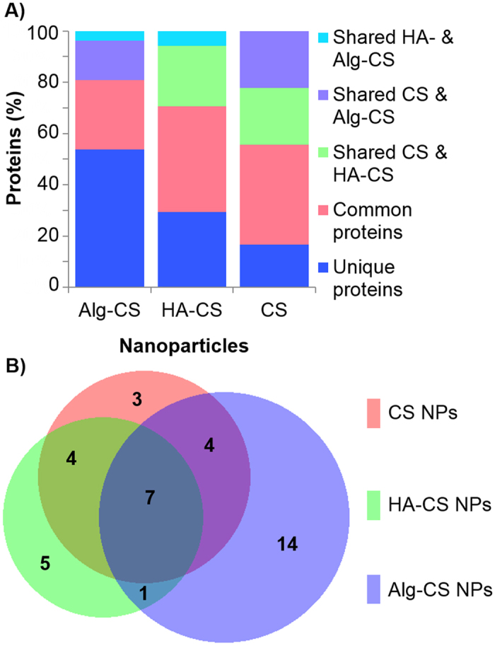
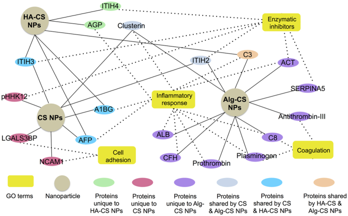
Studying the interactions of nanoparticles (NPs) with serum proteins is necessary for the rational development of nanocarriers. Optimum surface chemistry is a key consideration to modulate the formation of the serum protein corona (PC) and the resultant immune response. We investigated the constituent of the PC formed by hyaluronic acid-coated chitosan NPs (HA-CS NPs). Non-decorated chitosan NPs (CS NPs) and alginate-coated chitosan NPs (Alg-CS NPs) were utilized as controls. Results show that HA surface modifications significantly reduced protein adsorption relative to controls. Gene Ontology analysis demonstrates that HA-CS NPs were the least immunogenic nanocarriers. Indeed, less inflammatory proteins were adsorbed onto HA-CS NPs as opposed to CS and Alg-CS NPs. Interestingly, HA-CS NPs differentially adsorbed two unique anti-inflammatory proteins (ITIH4 and AGP), which were absent from the PC of both controls. On the other hand, CS and Alg-CS NPs selectively adsorbed a proinflammatory protein (Clusterin) that was not found on the surfaces of HA-CS NPs. While further studies are needed to investigate abilities of the PCs of only ITIH4 and AGP to modulate the interaction of NPs with the host immune system, our results suggest that this proof-of-concept could potentially be utilized to reduce the immunogenicity of a wide range of nanomaterials.


Conflict of interest statement

The authors declare that they have no competing interests.

Figures

Figure 1
Figure 1
Size and surface chemistry characterization of nanoparticles. (A) Size and (B) zeta potential distributions of chitosan nanoparticles (CS NPs), hyaluronic acid-coated CS NPs (HA-CS NPs), and alginate-coated CS NPs (Alg-CS NPs). (C) Statistical analysis and quantifications of size diameters and zeta potentials. Error bars represent three independent experiments.
Figure 2
Figure 2
Pattern and loading of protein coronas. (A) Size distributions of CS, HA-CS, and Alg-CS NPs before and after incubation with serum. (B) SDS-PAGE gel electrophoresis of chitosan nanoparticles (CS NPs), hyaluronic acid-coated CS NPs (HA-CS NPs), and alginate-coated CS NPs (Alg-CS NPs). 12% gel was used, and 4 µg of protein marker was loaded. (C) Bar graph depicting the quantitative differences in the amount of corona proteins (µg/µL) that were adsorbed to each of the different NPs. Error bars represent three independent experiments.
Figure 3
Figure 3
Mass spectrometric analysis. (A) Overlap comparison of protein coronas formed by chitosan nanoparticles (CS NPs), hyaluronic acid-coated CS NPs (HA-CS NPs), and alginate-coated CS NPs (Alg-CS NPs). (B) A Venn diagram depicting the differences and similarities in the number of the corona proteins identified between each nanoparticle.
Figure 4
Figure 4
Gene Ontology terms analysis. (A) Pie chart depicting the percentage of involvement of the identified proteins and their molecular functions. (B) A bar graph comparing the different molecular functions of proteins adsorbed to NPs.
Figure 5
Figure 5
Inflammatory focused nanoparticle-protein-function network. A network map of chitosan nanoparticles (CS NPs), hyaluronic acid-coated CS NPs (HA-CS NPs), and alginate-coated CS NPs (Alg-CS NPs) was analyzed that adsorb nineteen serum proteins involved in four inflammatory-related molecular functions. Spherical nodes in the map represent NPs, oval nodes represent proteins, while yellow rectangular nodes represent molecular functions. Oval nodes were color coded to distinguish unique proteins from those shared by two different NPs. Solid lines represent proteins absorbed onto NPs, while the dotted lines represent the molecular functions related to the identified proteins.
Figure 6
Figure 6
Proposed mechanism of creating immune modulating nanomaterials. Functionalizing of NPs with either anti-inflammatory proteins (AGP/ITIH4) or a proinflammatory protein (Clusterin) to produce immune suppressing/activating surfaces.

Similar articles

Cited by

References

    1. De Jong WH, Borm PJ. Drug delivery and nanoparticles:applications and hazards. International journal of nanomedicine. 2008;3:133–149. doi: 10.2147/IJN.S596. - DOI - PMC - PubMed
    1. Farokhzad OC, Langer R. Impact of nanotechnology on drug delivery. ACS nano. 2009;3:16–20. doi: 10.1021/nn900002m. - DOI - PubMed
    1. Lynch I, Dawson KA. Protein-nanoparticle interactions. Nano Today. 2008;3:40–47. doi: 10.1016/S1748-0132(08)70014-8. - DOI
    1. Walkey CD, Chan WCW. Understanding and controlling the interaction of nanomaterials with proteins in a physiological environment. Chemical Society Reviews. 2012;41:2780–2799. doi: 10.1039/C1CS15233E. - DOI - PubMed
    1. Monopoli MP, Aberg C, Salvati A, Dawson KA. Biomolecular coronas provide the biological identity of nanosized materials. Nature Nanotechnology. 2012;7:779–786. doi: 10.1038/nnano.2012.207. - DOI - PubMed

Publication types