. 2017:2017:3175748.
doi: 10.1155/2017/3175748. Epub 2017 Aug 10.

Different Angiogenic Potentials of Mesenchymal Stem Cells Derived from Umbilical Artery, Umbilical Vein, and Wharton's Jelly

Affiliations

Different Angiogenic Potentials of Mesenchymal Stem Cells Derived from Umbilical Artery, Umbilical Vein, and Wharton's Jelly

Lu Xu et al. Stem Cells Int. 2017.

Abstract

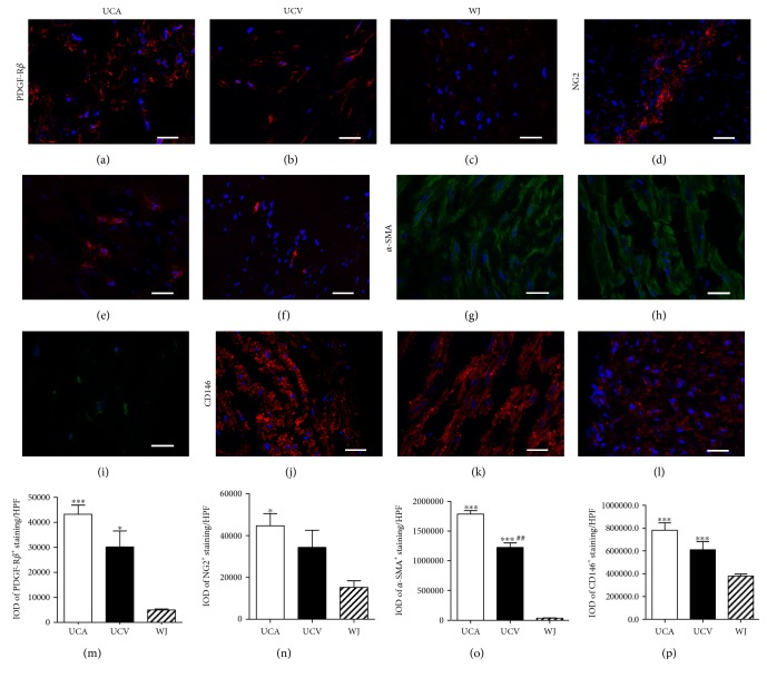
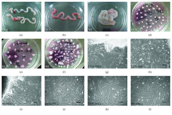
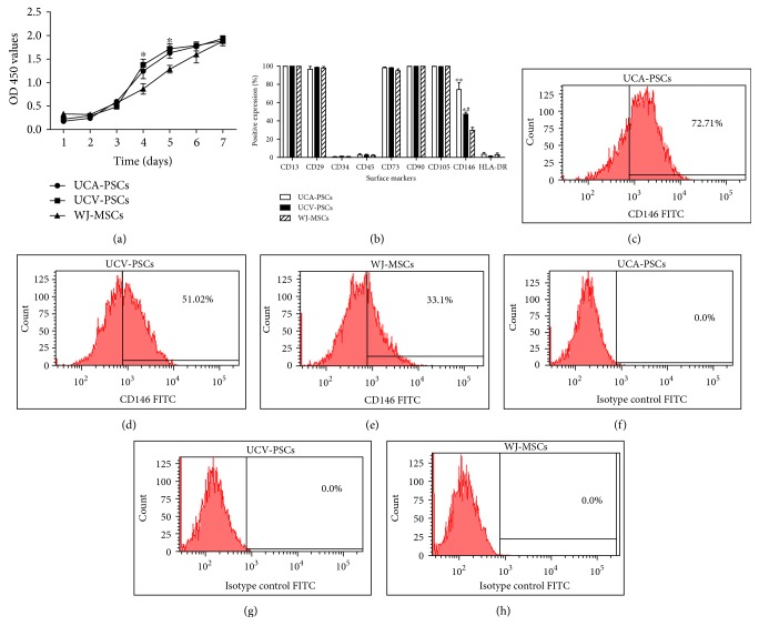
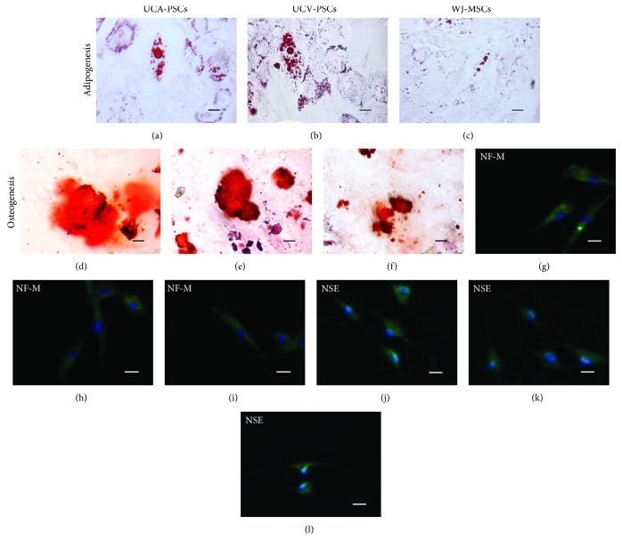
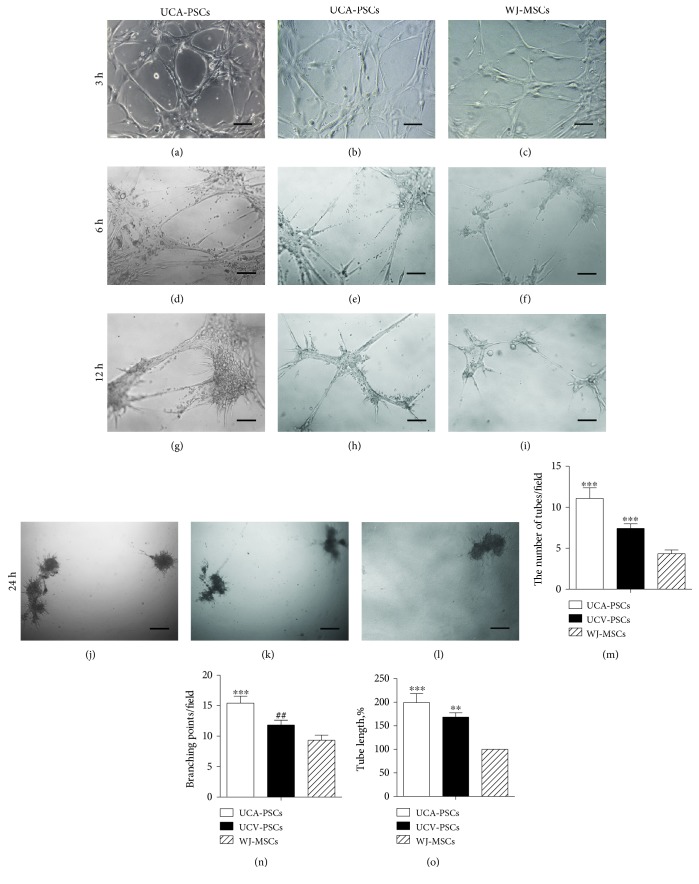
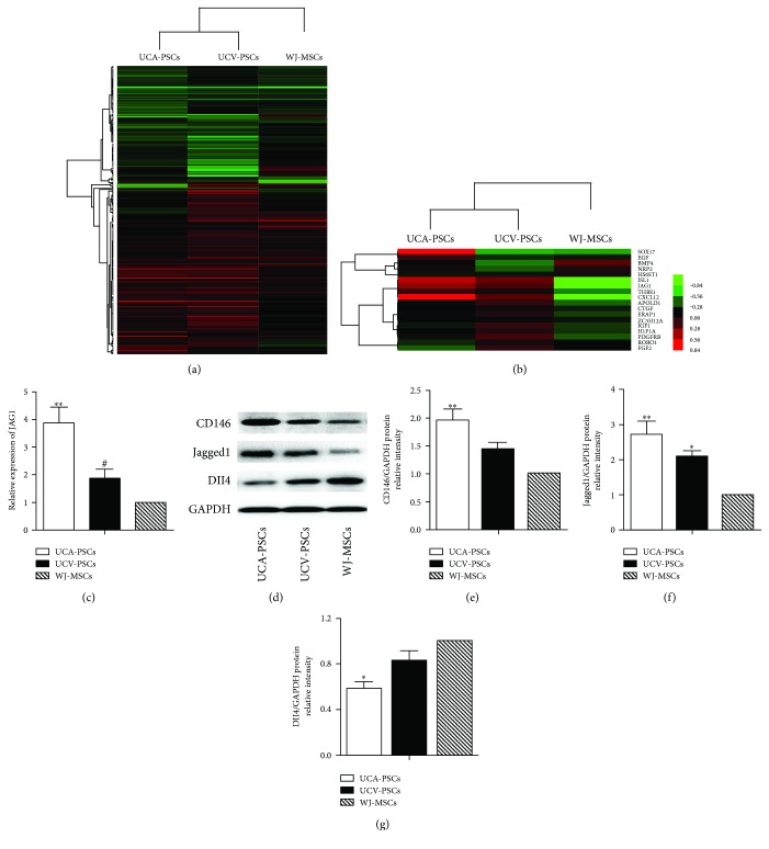
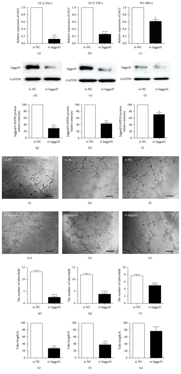
Human mesenchymal stem cells derived from the umbilical cord (UC) are a favorable source for allogeneic cell therapy. Here, we successfully isolated the stem cells derived from three different compartments of the human UC, including perivascular stem cells derived from umbilical arteries (UCA-PSCs), perivascular stem cells derived from umbilical vein (UCV-PSCs), and mesenchymal stem cells derived from Wharton's jelly (WJ-MSCs). These cells had the similar phenotype and differentiation potential toward adipocytes, osteoblasts, and neuron-like cells. However, UCA-PSCs and UCV-PSCs had more CD146+ cells than WJ-MSCs (P < 0.05). Tube formation assay in vitro showed the largest number of tube-like structures and branch points in UCA-PSCs among the three stem cells. Additionally, the total tube length in UCA-PSCs and UCV-PSCs was significantly longer than in WJ-MSCs (P < 0.01). Microarray, qRT-PCR, and Western blot analysis showed that UCA-PSCs had the highest expression of the Notch ligand Jagged1 (JAG1), which is crucial for blood vessel maturation. Knockdown of Jagged1 significantly impaired the angiogenesis in UCA-PSCs. In summary, UCA-PSCs are promising cell populations for clinical use in ischemic diseases.


Figures

Figure 1
Figure 1
Immunolocalization of PDGF-Rβ, NG2, α-SMA, and CD146 in the human umbilical cord. Fluorescence imaging revealed a high incidence of PDGF-Rβ-positive (a–c), NG2-positive (d–f), α-SMA-positive (g–i), and CD146-positive (j–l) cells in the perivascular region. Bar: 25 μm. The integrated optical density (IOD) values of positive staining in five randomly selected high power fields of view were counted. ∗∗∗P < 0.001, versus WJ-MSCs. P < 0.05, versus WJ-MSCs. ##P < 0.01, UCV-PSCs versus UCA-PSCs.
Figure 2
Figure 2
Isolation and characterization of umbilical cord artery perivascular stem cells (UCA-PSCs), umbilical cord vein perivascular stem cells (UCV-PSCs), and Wharton's jelly mesenchymal stem cells (WJ-MSCs). (a–c) Three different compartments in human umbilical cord: umbilical arteries (UCA) (a), umbilical vein (UCV) (b), and Wharton's jelly (WJ) (c). (d–f) Isolation of UCA-PSCs (d), UCV-PSCs (e), and WJ-MSCs in the human umbilical cord (f). (g–i) Cells from the third passage showed similar fibroblastic morphology. Bar: 100 μm.
Figure 3
Figure 3
Proliferation and phenotype profile of umbilical cord artery perivascular stem cells (UCA-PSCs), umbilical cord vein perivascular stem cells (UCV-PSCs), and Wharton's jelly mesenchymal stem cells (WJ-MSCs). (a) Cell proliferation was continuously monitored for 7 days using cell-counting kit-8 (CCK-8), which showed that the number of UCA-PSCs was significantly higher than that of UCV-PSCs at days 4 and 5. However, the cell growth rate of UCA-PSCs, UCV-PSCs, and WJ-MSCs did not significantly differ. Bars represent the means ± SE of three independent experiments performed in triplicate. P < 0.05, UCV-PSCs versus UCA-PSCs. (b) Profiles of cell surface epitopes in UCA-PSCs, UCV-PSCs, and WJ-MSCs. Abundance of cells positive for CD13, CD29, CD34, CD45, CD73, CD90, CD105, CD146, and HLA-DR, expressed as percentages, in UCA-PSCs, UCV-PSCs, and WJ-MSCs. Bars represent the means ± SE of donor samples (n = 3). ∗∗P < 0.01, UCA-PSCs versus WJ-MSCs. P < 0.05, UCV-PSCs versus WJ-MSCs. #P < 0.05, UCV-PSCs versus UCA-PSCs. (c–h) Representative flow cytometric plots, including isotype control (IgG-FITC) (f–h).
Figure 4
Figure 4
Differentiation of umbilical cord artery perivascular stem cells (UCA-PSCs), umbilical cord vein perivascular stem cells (UCV-PSCs), and Wharton's jelly mesenchymal stem cells (WJ-MSCs). (a–c) For adipogenic differentiation, the cells were cultured in adipogenic induction medium for 14 days. The formation of lipid droplets was confirmed by oil red O staining. (d–f) Cells were cultured in osteogenesis induction medium for 21 days. Calcium deposition was confirmed by alizarin red staining. (g–l) Differentiation of cells to neuronal lineage after 18 h preinduction and 36 h induction was confirmed by immunofluorescence staining of neurofilament medium polypeptide (g–i) and neuron-specific enolase (j–l). Bar: 10 μm.
Figure 5
Figure 5
In vitro Matrigel tube formation assay. Umbilical cord artery perivascular stem cells (UCA-PSCs), umbilical cord vein perivascular stem cells (UCV-PSCs), and Wharton's jelly mesenchymal stem cells (WJ-MSCs). Cells (1 × 104/well) were treated with a low concentration of glucose and plated on Matrigel in 96-well tissue culture plates. Tube formation was microscopically compared after 3 h (a–c), 6 h (d–f), 12 h (g–i), and 24 h (j–l). Bar: 60 μm. The number of tubes (m) and branching point per field (n) and the total length of tubes per field (o) were quantified 3 h after treatment by counting 3–5 random fields/well under the microscope (magnification: 100x). UCA-PSCs and UCV-PSCs, especially UCA-PSCs, displayed greater tube formation ability than WJ-MSCs. n = 5. ∗∗∗P < 0.001, versus WJ-MSCs. ∗∗P < 0.01, UCV-PSCs versus WJ-MSCs. ##P < 0.01, UCV-PSCs versus UCA-PSCs.
Figure 6
Figure 6
Expression of angiogenesis-related genes in the umbilical cord artery perivascular stem cells (UCA-PSCs), umbilical cord vein perivascular stem cells (UCV-PSCs), and Wharton's jelly mesenchymal stem cells (WJ-MSCs). (a) Comparison of gene expression patterns in UCA-PSCs, UCV-PSCs, and WJ-MSCs using microarray analysis. Red indicates upregulated genes while green indicates downregulated genes. (b) Gene expression heatmaps showed fold changes in the expression of a selection of genes involved in angiogenesis. (c) mRNA levels of Jagged1 in UCA-PSCs, UCV-PSCs, and WJ-MSCs measured by qRT-PCR. ∗∗P < 0.01, UCA-PSCs versus WJ-MSCs. #P < 0.05, UCV-PSCs versus UCA-PSCs. (d) Western blot analysis revealed that the CD146 and Jagged1 expression levels were the highest in UCA-PSCs, followed by UCV-PSCs and WJ-MSCs. While the Delta-like ligand (Dll4) protein displayed the opposite expression pattern in these three cell types. Quantitative analysis of CD146 (e), Jagged1 (f), and Dll4 (g) protein expression levels in UCA-PSCs, UCV-PSCs, and WJ-MSCs. Data are representative of three independent experiments performed in triplicate, and the expression of targeted protein was relative to the expression of GAPDH protein. ∗∗P < 0.01, versus WJ-MSCs. #P < 0.01, UCV-PSCs versus UCA-PSCs. P < 0.05, versus WJ-MSCs.
Figure 7
Figure 7
Knockdown of Jagged1 decreased tubule-like structures formation in the umbilical cord artery perivascular stem cells (UCA-PSCs), umbilical cord vein perivascular stem cells (UCV-PSCs), and Wharton's jelly mesenchymal stem cells (WJ-MSCs). (a–i) Jagged1 knockdown was effective. qRT-PCR analysis of Jagged1 mRNA levels in UCA-PSCs (a), UCV-PSCs (b), and WJ-MSCs (c) 48 h after the transfection with negative control siRNA (si-NC) or Jagged1 siRNA (si-Jagged1). ∗∗P < 0.01, si-Jagged1 versus si-NC; ∗∗∗P < 0.001, si-Jagged1 versus si-NC; P < 0.05, si-Jagged1 versus si-NC. Western blot analysis of Jagged1 expression in UCA-PSCs (d), UCV-PSCs (e), and WJ-MSCs (f) 72 h after the transfection with si-NC or si-Jagged1. Quantitative analysis of Jagged1 protein expression levels in UCA-PSCs (g), UCV-PSCs (h), and WJ-MSCs (i) 72 h after the transfection with si-NC or si-Jagged1. Data are representative of three independent experiments performed in triplicate, and the expression of targeted protein was relative to the expression of GAPDH protein. ∗∗P < 0.01, si-Jagged1 versus si-NC; P < 0.05, si-Jagged1 versus si-NC. Knockdown of Jagged1 decreased angiogenesis in UCA-PSCs, UCV-PSCs, and WJ-MSCs in vitro. (j–o) Representative images of tubular structures. Bar: 50 μm. Tube formation assays were performed 72 h after transfection of si-NC or si-Jagged1. The number of tubes in UCA-PSCs (p), UCV-PSCs (q), and WJ-MSCs (r), together with the total length of tubes per field in UCA-PSCs (s), UCV-PSCs (t), and WJ-MSCs (u) were quantified 3 h after treatment by counting 3–5 random fields/well under the microscope (magnification: 100x). n = 5. ∗∗∗P < 0.001, si-Jagged1 versus si-NC; ∗∗P < 0.01, si-Jagged1 versus si-NC.

Similar articles

Cited by

References

    1. Bronckaers A., Hilkens P., Martens W., et al. Mesenchymal stem/stromal cells as a pharmacological and therapeutic approach to accelerate angiogenesis. Pharmacology & Therapeutics. 2014;143(2):181–196. doi: 10.1016/j.pharmthera.2014.02.013. - DOI - PubMed
    1. Dominici M. L., Le Blanc K., Mueller I., et al. Minimal criteria for defining multipotent mesenchymal stromal cells. The International Society for Cellular Therapy position statement. Cytotherapy. 2006;8(4):315–317. doi: 10.1080/14653240600855905. - DOI - PubMed
    1. Pittenger M. F., Mackay A. M., Beck S. C., et al. Multilineage potential of adult human mesenchymal stem cells. Science. 1999;284(5411):143–147. doi: 10.1126/science.284.5411.143. - DOI - PubMed
    1. Teixeira F. G., Carvalho M. M., Sousa N., Salgado A. J. Mesenchymal stem cells secretome: a new paradigm for central nervous system regeneration. Cellular and Molecular Life Sciences. 2013;70(20):3871–3882. - PMC - PubMed
    1. Carvalho M. M., Teixeira F. G., Reis R. L., Sousa N., Salgado A. J. Mesenchymal stem cells in the umbilical cord: phenotypic characterization, secretome and applications in central nervous system regenerative medicine. Current Stem Cell Research & Therapy. 2011;6(3):221–228. - PubMed

LinkOut - more resources