Review
. 2017:2017:7941563.
doi: 10.1155/2017/7941563. Epub 2017 Aug 9.

Aging: Molecular Pathways and Implications on the Cardiovascular System

Affiliations
Review

Aging: Molecular Pathways and Implications on the Cardiovascular System

Arthur José Pontes Oliveira de Almeida et al. Oxid Med Cell Longev. 2017.

Abstract

The world's population over 60 years is growing rapidly, reaching 22% of the global population in the next decades. Despite the increase in global longevity, individual healthspan needs to follow this growth. Several diseases have their prevalence increased by age, such as cardiovascular diseases, the leading cause of morbidity and mortality worldwide. Understanding the aging biology mechanisms is fundamental to the pursuit of cardiovascular health. In this way, aging is characterized by a gradual decline in physiological functions, involving the increased number in senescent cells into the body. Several pathways lead to senescence, including oxidative stress and persistent inflammation, as well as energy failure such as mitochondrial dysfunction and deregulated autophagy, being ROS, AMPK, SIRTs, mTOR, IGF-1, and p53 key regulators of the metabolic control, connecting aging to the pathways which drive towards diseases. In addition, senescence can be induced by cellular replication, which resulted from telomere shortening. Taken together, it is possible to draw a common pathway unifying aging to cardiovascular diseases, and the central point of this process, senescence, can be the target for new therapies, which may result in the healthspan matching the lifespan.


Figures

Figure 1
Figure 1
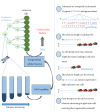
Aging and health. (a) The global population will increase from 12% in 2015 to almost 22% in 2050 [1]. (b) Despite the increase in lifespan, the individual healthspan does not follow this growth, which means that targeting aging with new therapies is essential to minimize the onset of aging-related diseases. (c) At the cellular level, aging is characterized by an increase of senescent cells in the organism, caused by several factors, including oxidative stress, systemic inflammation, mitochondrial dysfunction, deregulated nutrient sensitivity, autophagy dysfunction, and telomere shortening. The same mechanisms that lead to aging drive towards age-related diseases, in particular, the cardiovascular diseases, the major cause of death in the worldwide.
Figure 2
Figure 2
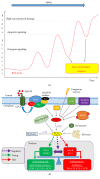
Senescence and aging. Aging is characterized by senescent cell accumulation into the body. Senescence can be achieved replicatively or induced by stress. Once activated, the p16 and p53/p21 pathways converge with each other, regulating the Rb mechanism, leading to cell cycle arrest, and consequently, the senescence. This results in the release of cytokines and chemokines, driving towards a systemic inflammatory condition that lead to aging and age-related diseases. The senescent cells are characterized by a high lysosomal β-galactosidase activity and, in association with others characteristic factors, consist the gold standard for the senescence characterization.
Figure 3
Figure 3
Role and function of telomeres in DNA protection. After each cell division, each chromosome loses a part of its telomeres, a region characterized by thousands of repeated sequences of nitrogenous bases. At a critical point, cells with shortened telomeres stop to divide, leading to senescence and resulting in aging and CVDs. Cells with high replicative rates such as stem cell lineages express telomerase, an enzyme capable of reversing telomere shortening. This enzyme plays a key role in the development of new therapies that aim to slow or reverse the aging process.
Figure 4
Figure 4
Redox potential controls cell fate. One of the hallmarks of aging is the increase in ROS levels production. New approaches define this increase as a compensatory cellular response with the original purpose to maintain cellular homeostasis and, from a certain limit, as a factor that aggravates aging. (a) The increase in ROS levels, first as a factor that activates survival pathways, continues to increase as a consequence of the deficiency in the antioxidant system, generating other cellular responses such as apoptosis, with a failure in apoptotic signalling, and driving towards severe cellular damage, such as necrosis. (b) Several sources of ROS contribute to the increase of redox potential, a factor that shifts the balance to the transcription of pro-inflammatory factors, while the antioxidant genes are silenced, connecting ROS and inflammation to aging.
Figure 5
Figure 5
Schematic overview of nucleus-mitochondria communication in aging. Decreased SIRT1 activity due to decreased NAD+ levels leads to changes in various gene expression. (1) Decreased PGC-1α/β levels, leading to decline the mitochondria biogenesis. (2) Increased expression of HIF-α, leading to a pseudohypoxia state and, consequently, a missed nucleus-mitochondria communication, driving towards a failure in coding OXPHOS genes. (3) Increased expression of Nf-kB levels, leading to inflammation, plus decreasing NAMPT production, a precursor of NAD+. (4) Decreased expression of FOXO, a factor that participates in cytoprotection. These responses are accompanied by increased ROS levels, a factor that activates AMPK, acting together as factors that counteract the decrease of SIRT1 activity. Increasing the ROS levels from a certain limit promote DNA damage, creating a paradoxical effect. In the mitochondria, the failure in the OXPHOS leads to a decrease in energy supply and, combined with oxidative stress, drive towards mitochondrial dysfunction. These factors combined lead to cellular stress and consequently to senescence. The interaction of oxidative stress and NAD+ levels are still unclear and may be an important source to understand how redox potential controls cellular energy metabolism.
Figure 6
Figure 6
Role of autophagy as cellular scavengers. Autophagy is mainly regulated by two energy sensors: mTOR and AMPK. mTOR is an inhibitor of autophagy and is activated when there are abundant cellular nutrients. AMPK is activated when nutrients deplete, inducing autophagy by inhibiting mTOR, as well as direct activation of autophagy. This mechanism is important for cell “cleaning,” degrading damaged organelles, protein aggregates, and other cellular toxic components. After the formation of the autophagosome, there is fusion with the lysosome, occurring the cleavage of the degraded material. There are two other types of autophagy: microautophagy, with direct involvement of the material by the lysosome. In addition, there is a chaperone-mediated autophagy, encompassing the material via the LAMP-2A receptor. Together, these mechanisms improve metabolism, being an energy source through recycling amino acids and, eventually, participating in cellular quality control, which promotes an improvement in the individual lifespan and healthspan.
Figure 7
Figure 7
Metabolic control involved in aging and on the cardiovascular system. IGF-1, mTOR, AMPK, SIRT1, p53, and ROS are key regulators in metabolic control. Many of these pathways are complex involving crosstalk between them, with many paradoxical effects. The stimulation of the IGF-1 pathway by insulin promotes PI3K/AKT pathway activation, which induces the exclusion of FOXO from the nucleus, inhibiting its function; in addition, IGF-1 activates eNOS, increasing NO availability, improving the vascular function. IGF-1 also activates the RAS/p38MAPK pathway inducing mechanisms of cell growth and proliferation. Finally, IGF-1 stimulates vesicles containing GLUT-4 to the cell membrane, promoting the uptake of glucose, the main cellular energy substrate. There are also other two glucose transporters that help in glucose uptake such as GLUT-1 and GLUT-3; the last one can be downregulated by p53 via Nf-kB. Under normal conditions, mostly, pyruvate is directed to the mitochondria, producing ATP by OXPHOS. In age, NAD+ levels decrease, driving towards a loss in SIRT1 activity, resulting in mitochondrial dysfunction via PGC-1α/β and HIF-α. Thus, pyruvate is directed to lactate production, even in the presence of O2, a process known as “the Warburg effect”. This metabolic shift is essential for increasing biomass, stimulating cell growth, proliferation, and differentiation, which promote angiogenesis. In this way, a mitochondrial dysfunction results in a decreased ATP production, activating AMPK. This protein stimulates autophagy, generating energy for the cell. In addition, it stimulates p53, which inhibit the uptake and conversion of glucose, to stimulate OXPHOS activity, generating an antiproliferative effect. Finally, ROS produced by mitochondrial dysfunction stimulates several signalling pathways, such as AMPK, but also activates AKT, which stimulates mTOR, being the redox potential the major regulator of this balance.
Figure 8
Figure 8
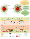
Young and aged vascular comparison in two different perspectives. In vascular aging, the remodelling occurs due to the accumulation of senescent and dysfunctional cells in response to the environmental changes caused by age. (a) In the aged vessel, there is a loss of the vessel elasticity, due to the raises of contracting factors, plus an increase in the number of muscle cells. These factors combined drive towards the matrix change, with inflammatory infiltrates and fibrosis, leading to vascular hypertrophy. (b) In young blood, there is a predominance of growth factors, in addition to healthy cells of immunity and progenitor cells driving towards vascular “cleaning” and regeneration, respectively. In the aged blood, it is checked that there is a predominance in proinflammatory factors, released largely by senescent cells. The senescent cells accumulate with age in response to a failure of the immune system, a term known as immunosenescence. In addition, there is an increase in fibroblast proliferation, leading to a stressful environment related to the vascular remodelling.

Similar articles

Cited by

References

    1. UN. HelpAge, Global AgeWatch Index 2015: Insight Report. London WC1A 9GB, UK: HelpAge International; 2015. UNDESA Population Division, World population prospects: the 2015 revision.
    1. WHO. NCD Mortality and Morbidity. World Health Organization; 2012. http://www.who.int/gho/ncd/mortality_morbidity/en/
    1. Campisi J. Aging, cellular senescence, and cancer. Annual Review of Physiology. 2013;75(1):685–705. doi: 10.1146/annurev-physiol-030212-183653. - DOI - PMC - PubMed
    1. López-Otín C., Blasco M. A., Partridge L., Serrano M., Kroemer G. The hallmarks of aging. Cell. 2013;153(6):1194–1217. doi: 10.1016/j.cell.2013.05.039. - DOI - PMC - PubMed
    1. Riera C. E., Merkwirth C., Filho C. D. D. M., Dillin A. Signaling networks determining life span. Annual Review of Biochemistry. 2016;85(1):35–64. doi: 10.1146/annurev-biochem-060815-014451. - DOI - PubMed