

Clinical impact of confocal laser endomicroscopy in the management of gastrointestinal lesions with an uncertain diagnosis
- PMID: 28874959
- PMCID: PMC5565504
- DOI: 10.4253/wjge.v9.i8.389
Clinical impact of confocal laser endomicroscopy in the management of gastrointestinal lesions with an uncertain diagnosis
Abstract
Aim: To evaluate the clinical impact of confocal laser endomicroscopy (CLE) in the diagnosis and management of patients with an uncertain diagnosis.
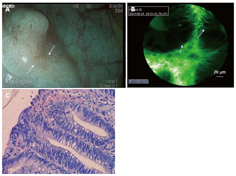
Methods: A retrospective chart review was performed. Patients who underwent CLE between November 2013 and October 2015 and exhibited a poor correlation between endoscopic and histological findings were included. Baseline characteristics, indications, previous diagnostic studies, findings at the time of CLE, clinical management and histological results were analyzed. Interventions based on CLE findings were also analyzed. We compared the diagnostic accuracy of CLE and target biopsies of surgical specimens.
Results: A total of 144 patients were included. Of these, 51% (74/144) were female. The mean age was 51 years old. In all, 41/144 (28.4%) lesions were neoplastic (13 bile duct, 10 gastric, 8 esophageal, 6 colonic, 1 duodenal, 1 rectal, 1 ampulloma and 1 pancreatic). The sensitivity, specificity, positive predictive value, negative predictive value, and observed agreement when CLE was used to detect N-lesions were 85.37%, 87.38%, 72.92%, 93.75% and 86.81%, respectively. Cohen's Kappa was 69.20%, thus indicating good agreement. Changes in management were observed in 54% of the cases.
Conclusion: CLE is a new diagnostic tool that has a significant clinical impact on the diagnosis and treatment of patients with uncertain diagnosis.
Keywords: Barret esophagus; Biliary strictures; Confocal laser endomicroscopy; Gastrointestinal cancer; In vivo microscopy; Pancreatic cyst.
Conflict of interest statement
Conflict-of-interest statement: The authors have no conflict of interests.
Figures

References
-
- Bartels F, Hahn HJ, Stolte M, Schmidt-Wilcke HA. [Quality of diagnostic procedures and frequency of endoscopically defined diseases of the upper gastrointestinal tract] Z Gastroenterol. 2003;41:311–318. - PubMed
-
- Isaacs KL. Upper gastrointestinal tract endoscopy in inflammatory bowel disease. Gastrointest Endosc Clin N Am. 2002;12:451–462, vii. - PubMed
-
- Sharma P, Hawes RH, Bansal A, Gupta N, Curvers W, Rastogi A, Singh M, Hall M, Mathur SC, Wani SB, et al. Standard endoscopy with random biopsies versus narrow band imaging targeted biopsies in Barrett’s oesophagus: a prospective, international, randomised controlled trial. Gut. 2013;62:15–21. - PubMed
-
- Calhoun BC, Gomes F, Robert ME, Jain D. Sampling error in the standard evaluation of endoscopic colonic biopsies. Am J Surg Pathol. 2003;27:254–257. - PubMed
LinkOut - more resources
Full Text Sources
Other Literature Sources
