

doi: 10.1007/s13238-017-0456-9.
Cryo-EM structure of Mycobacterium smegmatis ribosome reveals two unidentified ribosomal proteins close to the functional centers
Affiliations
- PMID: 28875450
- PMCID: PMC5876184
- DOI: 10.1007/s13238-017-0456-9
Item in Clipboard
Cryo-EM structure of Mycobacterium smegmatis ribosome reveals two unidentified ribosomal proteins close to the functional centers
Protein Cell.
2018 Apr.
No abstract available
Figures
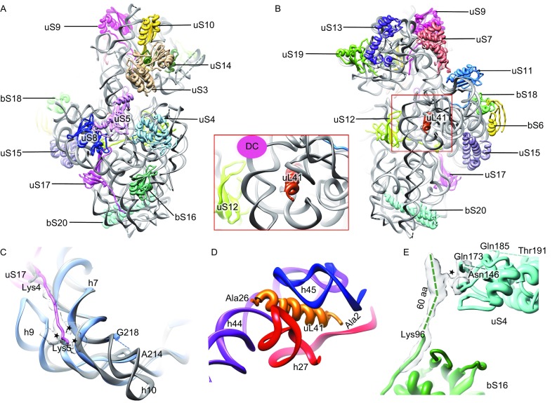
Overall atomic model and unique features of the
MS
30S. (A and B) Atomic model of the MS30S, viewed from the solvent surface (A) and intersubunit surface (B). Inset, zoom-in view of uL41 close to the decoding center (DC). (C) The extended N-terminus of uS17 interacts with h7 and h9. Counterparts in the EC70S are colored gray. The coordinates of the E. coli components are from a crystallography study (PDB 4KIY) (Pulk and Cate, 2013). (D) Ribosomal protein uL41 is surrounded by h27, h44, and h45. (E) The extended C-terminus of bS16 interacts with globular domain of uS4. The interaction sites are indicated by asterisks
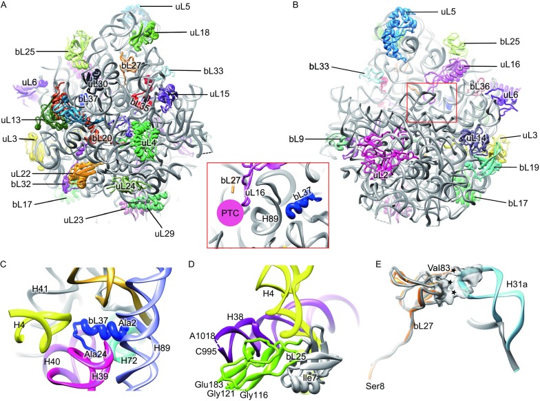
Overall atomic model and unique features in the
MS
50S. (A and B) Atomic model of the MS50S, viewed from solvent surface (A) and inter-subunit surface (B). Inset, relative orientation of bL27 with respect to the peptidyl transferase center (PTC). (C) Ribosomal protein bL37 is encircled by H4, H39, H40, base of H41 and H42, H72 and H89. (D) The extra C-terminal domain of bL25 is in proximity to H38 (purple). Conserved N-terminal domain of bL25 is colored gray while the extra C-terminal domain is colored green. (E) The extended H31 interacts with bL27, N-terminus of which extends to the peptidyl transfer center. The interaction sites are indicated by asterisks. Protein bL27 and H31a in the MS70S are colored with orange and cyan, respectively, while their counterparts in the EC70S are in gray. The coordinates of the E. coli components are from a crystallography study (PDB 4KIX) (Pulk and Cate, 2013)
References
Publication types
MeSH terms
Substances
LinkOut - more resources
Full Text Sources
Other Literature Sources
