. 2018 Jan 1;142(1):81-91.
doi: 10.1002/ijc.31029. Epub 2017 Sep 23.

The contribution of toll-like receptor signaling to the development of liver fibrosis and cancer in hepatocyte-specific TAK1-deleted mice

Affiliations

The contribution of toll-like receptor signaling to the development of liver fibrosis and cancer in hepatocyte-specific TAK1-deleted mice

Isabelle Jingyi Song et al. Int J Cancer. .

Abstract

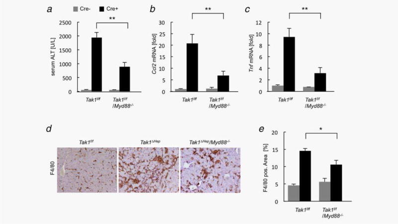
Hepatocyte death is associated with liver inflammation, fibrosis and hepatocellular carcinoma (HCC). Damaged cells trigger inflammation through activation of Toll-like receptors (TLRs). Although the role of TLR4 in HCC development has been reported, the role of TLR9 in the development of HCC remains elusive. To investigate the role of TLR4 and TLR9 signaling in liver inflammation-fibrosis-cancer axis, we took advantage of mice with hepatic deletion of transforming growth factor-β-activated kinase 1 (Tak1ΔHep) that develop spontaneous liver injury, inflammation, fibrosis, and HCC, recapitulating the pathology of human HCC. We generated double knockout mice lacking genes of our interest with hepatic Tak1. Tak1ΔHep mice and Tlr4-deficient Tak1ΔHep mice had similar serum ALT levels, but Tlr4-deficient Tak1ΔHep mice exhibited significantly reduced macrophage infiltration, myofibroblast activation and tumor formation. Ablation of TLR9 reduced spontaneous liver injury, inflammation, fibrosis, and cancer development in Tak1ΔHep mice. In addition, the common adaptor, myeloid differentiation factor 88 (MyD88)-deficient Tak1ΔHep mice also attenuated liver injury, macrophage recruitment, collagen deposition, and tumor growth compared with control Tak1ΔHep mice. Genetic ablation of TNF receptor type I (TNFR) in Tak1ΔHep mice remarkably reduced liver inflammation-fibrosis-cancer axis. Surprisingly, disruption of interleukin-1 receptor (IL-1R) had no effect on liver injury and tumor formation, although Il1r-deficient Tak1ΔHep showed attenuated macrophage infiltration and collagen deposition. In conclusion, TLR4- and TLR9-MyD88 are driving forces of progression to HCC accompanied by liver inflammation and fibrosis in Tak1ΔHep mice. Importantly, TLR4 and TLR9 downstream TNFR, but not IL-1R signaling is crucial for the development of HCC in Tak1ΔHep mice.

Keywords: HCC; TAK1; TNF receptor type I; liver fibrosis; toll-like receptors.


Conflict of interest statement

Conflict of interest: The authors declare that they have no conflict of interest to declare.

Figures

Figure 1
Figure 1
Ablation of Tlr4 or Tlr9 in Tak1ΔHep mice attenuates spontaneous liver inflammation. (a) Serum ALT levels were measured in Tak1fl/fl, Tak1ΔHep, Tak1fl/fl/Tlr4−/−, Tak1ΔHep/Tlr4−/−, Tak1fl/fl/Tlr9−/− and Tak1ΔHep/Tlr9−/− mice at the age of 1 month (n = 8–18). (b, c) Hepatic messenger RNA expression of inflammatory genes (Ccl2 and Tnf) in liver tissues from 1-month-old mice were measured by qPCR. (d, e) Macrophage infiltration was determined by immunohistochemistry for F4/80 (d). F4/80 positive area was quantified by ImageJ software (e). Original magnification ×200. Gray bar, Alb-Cre negative mice. Black bar, Alb-Cre positive mice. Data represent mean ± SEM; **, p < 0.01; *, p < 0.05; n.s., not significant. [Color figure can be viewed at wileyonlinelibrary.com]
Figure 2
Figure 2
TLR4 and TLR9 signaling is required for spontaneous liver fibrosis in Tak1ΔHep mice. (a) α-smooth muscle actin (α-SMA) expression in the livers of Tak1fl/fl, Tak1ΔHep, Tak1ΔHep/Tlr4−/−, and Tak1ΔHep/Tlr9−/− mice at the age of 1 month was determined by immunohistochemistry (left) (n = 6–8). Fibrillar collagen deposition in the livers of mice at the age of 9 months was evaluated by Sirius red staining (right) (n = 4–6). Original magnification, ×200 for immunohistochemistry for α-SMA and ×100 for Sirius Red Staining. (b, c) Quantification for staining for α-SMA (b) and Sirius Red staining (c). (d, e) Hepatic messenger RNA expression of fibrogenic genes (Timp1 and Tgfb1) in liver tissues from 1-month-old mice was determined by qPCR. Gray bar, Alb-Cre negative mice. Black bar, Alb-Cre positive mice. Data represent mean ± SEM; **p < 0.01; *p < 0.05. [Color figure can be viewed at wileyonlinelibrary.com]
Figure 3
Figure 3
Additional deletion of Myd88 reduces spontaneous liver inflammation in the livers of Tak1ΔHep mice. (a) Serum ALT levels were measured in Tak1fl/fl, Tak1ΔHep, Tak1fl/fl/Myd88−/− and Tak1ΔHep/Myd88−/− mice at the age of 1 month (n = 4–17). (b, c) Hepatic messenger RNA expressions of Ccl2 (b) and Tnf (c) were assessed by qPCR. (d, e) Representative pictures (d) and quantification (e) of immunohistochemistry for F4/80. Original magnification ×200. Gray bar, Alb-Cre negative mice. Black bar, Alb-Cre positive mice. Data represent mean ± SEM; **p < 0.01; *, p < 0.05. [Color figure can be viewed at wileyonlinelibrary.com]
Figure 4
Figure 4
Loss of Myd88 in Tak1ΔHep mice suppresses spontaneous liver fibrosis. (a) Representative pictures of immunohistochemistry for α-SMA (left) in the livers of Tak1fl/fl, Tak1ΔHep, and Tak1ΔHep/Myd88−/− mice at the age of 1 month (n = 4–7). Sirius Red staining (right) in the livers of mice at the age of 9 months (n = 5–6). Original magnification, ×200 for immunohistochemistry for α-SMA and ×100 for Sirius Red staining. (b, c) Quantification for staining for α-SMA (b) and Sirius Red staining (c). (d, e) Hepatic messenger RNA expressions of Timp1 (d) and Tgfb1 (e) in liver tissues from 1-month-old mice were assessed by qPCR. Gray bar, Alb-Cre negative mice. Black bar, Alb-Cre positive mice. Data represent mean ± SEM; **P < 0.01; *P < 0.05. [Color figure can be viewed at wileyonlinelibrary.com]
Figure 5
Figure 5
TNF receptor and IL-1 receptor signaling is partially responsible for macrophage infiltration and liver fibrosis in Tak1ΔHep mice. (a) Serum ALT levels of 1-month-old Tak1fl/fl, Tak1ΔHep, Tak1fl/fl/Tnfr−/−, Tak1ΔHep/Tnfr−/−, Tak1fl/fl/Il1r−/−, and Tak1ΔHep/Il1r−/− mice (n = 5–17). (b, c) Hepatic messenger RNA expression of Ccl2 (b) and Timp1 (c) was measured by qPCR in 1-month-old mice. (d, e) Representative pictures (d) and quantification (e) of immunohistochemistry for F4/80 and α-SMA (1-month-old, n = 4–10), and Sirius Red staining (9-month-old, n = 4–6). Original magnification, ×200 for immunohistochemistry for F4/80 and α-SMA, and ×100 for Sirius Red staining. Gray bar, Alb-Cre negative mice. Black bar, Alb-Cre positive mice. Data represent mean ± SEM; **p < 0.01; *p < 0.05; n.s., not significant. [Color figure can be viewed at wileyonlinelibrary.com]
Figure 6
Figure 6
Spontaneous hepatocarcinogenesis in Tak1ΔHep mice is suppressed by additional deletion of TLR4, TLR9, MyD88, and TNFR, but not IL-1R. (a) Representative macroscopic pictures of livers of 9-month-old Tak1ΔHep, Tak1ΔHep/Tlr4−/−, Tak1ΔHep/Tlr9−/−, Tak1ΔHep/Myd88−/−, Tak1ΔHep/Tnfr−/−, and Tak1ΔHep/Il1r−/− mice. (b) The number of tumor nodules per mouse was counted (left) and the maximal tumor size was measured (right). (n = 8–28). **p < 0.01; *p < 0.05; n.s., not significant. [Color figure can be viewed at wileyonlinelibrary.com]

References

    1. Roh YS, Song J, Seki E. TAK1 regulates hepatic cell survival and carcinogenesis. J Gastroenterol. 2014;49:185–94. - PMC - PubMed
    1. Shim JH, Xiao C, Paschal AE, et al. TAK1, but not TAB1 or TAB2, plays an essential role in multiple signaling pathways in vivo. Genes Dev. 2005;19:2668–81. - PMC - PubMed
    1. Sato S, Sanjo H, Takeda K, et al. Essential function for the kinase TAK1 in innate and adaptive immune responses. Nat Immunol. 2005;6:1087–95. - PubMed
    1. Tang M, Wei X, Guo Y, et al. TAK1 is required for the survival of hematopoietic cells and hepatocytes in mice. J Exp Med. 2008;205:1611–9. - PMC - PubMed
    1. Inokuchi S, Aoyama T, Miura K, et al. Disruption of TAK1 in hepatocytes causes hepatic injury, inflammation, fibrosis, and carcinogenesis. Proc Natl Acad Sci U S A. 2010;107:844–9. - PMC - PubMed

Publication types

MeSH terms