

Redo surgery using IntraLase femtosecond laser for treating a decentered laser in situ keratomileusis flap
- PMID: 28875760
- PMCID: PMC5971497
- DOI: 10.1177/0300060517718989
Redo surgery using IntraLase femtosecond laser for treating a decentered laser in situ keratomileusis flap
Abstract
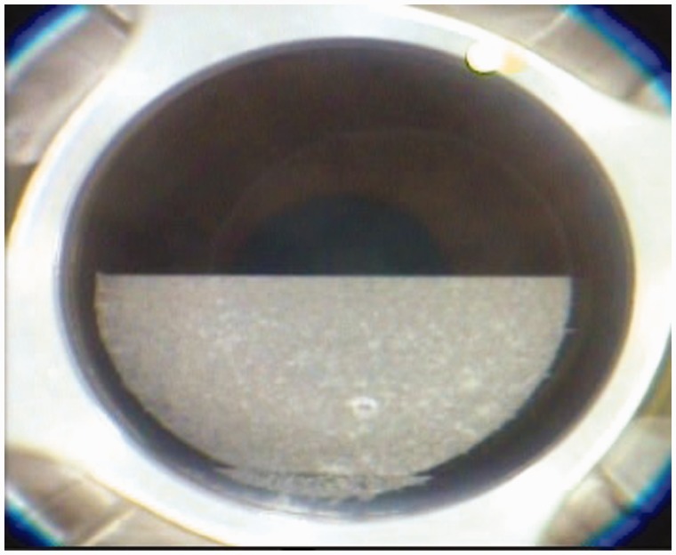
Objective Decentered flaps are rarely reported after femtosecond laser-assisted in situ keratomileusis flap procedures. We present a patient with a decentered flap after preparation of a corneal flap using the Femto LDV technique. Methods The 22-year-old man required a redo operation because of a decentered corneal flap. It was performed the same day at the patient's insistence and with his consent. The new corneal flap for the redo surgery was prepared using the femtosecond laser technique and IntraLase. Results Uncorrected visual acuity for each eye was 1.2 during the 12-month follow-up. The results of the Femtosecond laser technique showed good predictability and repeatability regarding the preparation of corneal flaps, but it still may cause some intraoperative complications. Conclusion Once redo surgery is needed, the size and depth of the initially prepared flap should be determined using anterior segment optical coherence tomography to pre-set the parameters for preparation of the redo flap.
Keywords: Decentered flap; Femto LDV; LASIK; optical coherence tomography (OCT); redo surgery; second flap.
Figures


References
-
- Durrie DS, Kezirian GM. Femtosecond laser versus mechanical keratome flaps in wavefront-guided laser in situ keratomileusis: prospective contralateral eye study. J Cataract Refract Surg 2005; 31: 120–126. DOI: 10.1016/j.jcrs.2004.09.046. - PubMed
-
- Nordan LT, Slade SG, Baker RN, et al. Femtosecond laser flap creation for laser in situ keratomileusis: six-month follow-up of initial U.S. clinical series. J Refract Surg 2003; 19: 8–14. - PubMed
-
- Muñoz G, Albarrán-Diego C, Sakla HF, et al. Transient light-sensitivity syndrome after laser in situ keratomileusis with the femtosecond laser incidence and prevention. J Cataract Refract Surg 2006; 32: 2075–2079. DOI: 10.1016/j.jcrs.2006.07.024. - PubMed
-
- Lifshitz T, Levy J, Klemperer I, et al. Anterior chamber gas bubbles after corneal flap creation with a femtosecond laser. J Cataract Refract Surg 2005; 31: 2227–2229. DOI: 10.1016/j.jcrs.2004.12.069. - PubMed
Publication types
MeSH terms
LinkOut - more resources
Full Text Sources
Other Literature Sources
