Review
. 2017 Sep 6;12(9):845-855.
doi: 10.1038/nnano.2017.167.

Multiscale technologies for treatment of ischemic cardiomyopathy

Affiliations
Review

Multiscale technologies for treatment of ischemic cardiomyopathy

Morteza Mahmoudi et al. Nat Nanotechnol. .

Abstract

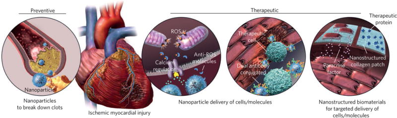
The adult mammalian heart possesses only limited capacity for innate regeneration and the response to severe injury is dominated by the formation of scar tissue. Current therapy to replace damaged cardiac tissue is limited to cardiac transplantation and thus many patients suffer progressive decay in the heart's pumping capacity to the point of heart failure. Nanostructured systems have the potential to revolutionize both preventive and therapeutic approaches for treating cardiovascular disease. Here, we outline recent advancements in nanotechnology that could be exploited to overcome the major obstacles in the prevention of and therapy for heart disease. We also discuss emerging trends in nanotechnology affecting the cardiovascular field that may offer new hope for patients suffering massive heart attacks.


Conflict of interest statement

Competing financial interests

R.L. and O.C.F. declare financial interests in Selecta Biosciences, Tarveda Therapeutics and Placon Therapeutics. R.L. declares financial interests in Moderna.

Figures

Figure 1
Figure 1. Applications of various nanoplatforms in the prevention and treatment of cardiovascular disease
Nanoplatforms can target and break down coronary artery plaques and prevent injuries caused by stenosis or occlusion of arteries. Nanoparticulate systems can also reduce the adverse effects of reperfusion injuries and regenerate/salvage myocardium after MI, through sustained and targeted delivery of cells, biomolecules and paracrine factors.
Figure 2
Figure 2. Dual-antibody-conjugated magnetic nanoparticles target therapeutic cells and regenerate the injured myocardium
a,b, Schematic showing the injection of the magnetic nanoparticles (NPs) with and without conjugated antibodies in vivo. c, Fluorescent imaging of the rat’s organs after injection of the therapeutic cells (rat bone marrow mononuclear cells labelled with a fluorescent dye) showing their targeted accumulation in the hearts of animals that received the dual-antibody-conjugated magnetic nanoparticles, unlike those that received regular magnetic nanoparticles. There are significantly fewer off-targeted therapeutic cells (for example, in lung) in animals that received dual-antibody-conjugated magnetic nanoparticles compared with the regular magnetic nanoparticles. d, Image of the whole heart section (trichrome staining) showing the significant reduction in scar size (blue) and substantial enhancement of viable (red) tissues in animals that received dual-antibody-conjugated magnetic nanoparticles compared with the regular magnetic nanoparticles and control sample (no therapeutic cells), 4 weeks post reperfusion. e, Echocardiography results, 4 weeks post reperfusion, revealed significant improvement in left ventricular ejection fraction (LVEF) in the targeted-nanoparticle group compared with the regular nanoparticle and control groups. f, Confocal microscopic images showing a significant effect of nanoparticle-targeted cell therapies on angiogenesis compared with the regular nanoparticle and control groups. Blue represents DAPI for cell nuclei and green is alpha smooth muscle actin (αSMA) for smooth muscle cells. Figure adapted from ref., Macmillan Publishers Ltd.
Figure 3
Figure 3. The use of a living contrast agent, MEs derived from magnetotactic bacteria, for safe labelling and precise monitoring of CMs
a, Transmission electron microscopy image of the magnetosome structure. b, Fluorescent images of the unlabelled (left panel) and ME-labelled (right panel) iPSC-derived chemically defined CMs demonstrating internalization of MEs in the CMs; red, green and blue colours show the MEs, cell membrane and cell nucleus, respectively. c, Propidium iodide (PI) viability assay demonstrates the safe labelling of the MEs (left and right panels show unlabelled and ME-labelled CMs, respectively). Blue colour shows the cell nucleus; red colour denotes dead cells. d,e, Intravascular (IV; d) and intramyocardial (IM; e) injection of the MEs showing no signs of infection or blood toxicity, as there were no substantial changes in the relative concentrations of liver and spleen enzymes. a.u., arbitrary units. f, Representative whole-body bioluminescence images of mice that received the phosphate-buffered saline control and CMs labelled either with ME or the magnetic nanoparticles (Molday) after 7 d. g, Bioluminescence images of the mice 14 d after cell injection (top) and corresponding in vivo MRI images of the murine hearts (bottom); the signal from the injected labelled cells is denoted by red arrows. h, Representative bioluminescence images of mice with dead CMs at day 14 of treatment with ME-labelled cells and the corresponding in vivo MRI images. i, Comparison of signal intensity for Molday (dark signal) and ME (no signal), showing significantly lower signal of ME*-labelled dead cells compared with other samples with positive signal (P < 0.05). Figure adapted from ref. , Macmillan Publishers Ltd.
Figure 4
Figure 4. Application of nanostructured cardiac patch device in repair/regeneration of MI
a, Different scenarios by which the engineered nanostructured scaffold can contribute to myocardial tissue repair following MI: (i) inhibition of adverse cardiac remodelling processes post-MI via physical mechanical support; (ii) targeted delivery and sustained release of anti-apoptotic factors; (iii) promoting angiogenesis (via delivery of angiogenic factors); and (iv) promoting proliferation of pre-existing native CMs (for example, through epicardial delivery of human FSTL1 peptide). b, Schematic demonstration of cellular and molecular processes in healthy (top), post-MI (middle) and patch-treated (bottom) heart tissue. The bottom panel delineates the role of a nanostructured cardiac patch in regenerating the damaged myocardium by inducing cell cycle re-entry among pre-existing CMs.

References

    1. Bui AL, Horwich TB, Fonarow GC. Epidemiology and risk profile of heart failure. Nat Rev Cardiol. 2011;8:30–41. - PMC - PubMed
    1. Johnson NB, et al. CDC National Health Report: leading causes of morbidity and mortality and associated behavioral risk and protective factors—United States, 2005–2013. MMWR Surveill Summ. 2014;63:3–27. - PubMed
    1. Thygesen K, Alpert JS, White HD. Universal definition of myocardial infarction. J Am Coll Cardiol. 2007;50:2173–2195. - PubMed
    1. Cassar A, Holmes DR, Jr, Rihal CS, Gersh BJ. Chronic coronary artery disease: diagnosis and management. Mayo Clin Proc. 2009;84:1130–1146. - PMC - PubMed
    1. White HD, Chew DP. Acute myocardial infarction. Lancet. 2008;372:570–584. - PubMed

Publication types

MeSH terms