. 2019 Mar;10(2):146-161.
doi: 10.1080/21541248.2017.1356432. Epub 2017 Sep 29.

Impairments in age-dependent ubiquitin proteostasis and structural integrity of selective neurons by uncoupling Ran GTPase from the Ran-binding domain 3 of Ranbp2 and identification of novel mitochondrial isoforms of ubiquitin-conjugating enzyme E2I (ubc9) and Ranbp2

Affiliations

Impairments in age-dependent ubiquitin proteostasis and structural integrity of selective neurons by uncoupling Ran GTPase from the Ran-binding domain 3 of Ranbp2 and identification of novel mitochondrial isoforms of ubiquitin-conjugating enzyme E2I (ubc9) and Ranbp2

Hemangi Patil et al. Small GTPases. 2019 Mar.

Abstract

The Ran-binding protein 2 (Ranbp2/Nup358) is a cytoplasmic and peripheral nucleoporin comprised of 4 Ran-GTP-binding domains (RBDs) that are interspersed among diverse structural domains with multifunctional activities. Our prior studies found that the RBD2 and RBD3 of Ranbp2 control mitochondrial motility independently of Ran-GTP-binding in cultured cells, whereas loss of Ran-GTP-binding to RBD2 and RBD3 are essential to support cone photoreceptor development and the survival of mature retinal pigment epithelium (RPE) in mice. Here, we uncover that loss of Ran-GTP-binding to RBD3 alone promotes the robust age-dependent increase of ubiquitylated substrates and S1 subunit (Pmsd1) of the 19S cap of the proteasome in the retina and RPE and that such loss in RBD3 also compromises the structural integrity of the outer segment compartment of cone photoreceptors only and without affecting the viability of these neurons. We also found that the E2-ligase and partner of Ranbp2, ubc9, is localized prominently in the mitochondrial-rich ellipsoid compartment of photoreceptors, where Ranbp2 is also known to localize with and modulate the activity of mitochondrial proteins. However, the natures of Ranbp2 and ubc9 isoforms to the mitochondria are heretofore elusive. Subcellular fractionation, co-immunolocalization and immunoaffinity purification of Ranbp2 complexes show that novel isoforms of Ranbp2 and ubc9 with molecular masses distinct from the large Ranbp2 and unmodified ubc9 isoforms localize specifically to the mitochondrial fraction or associate with mitochondrial components, whereas unmodified and SUMOylated Ran GTPase are excluded from the mitochondrial fraction. Further, liposome-mediated intracellular delivery of an antibody against a domain shared by the mitochondrial and nuclear pore isoforms of Ranbp2 causes the profound fragmentation of mitochondria and their delocalization from Ranbp2 and without affecting Ranbp2 localization at the nuclear pores. Collectively, the data support that Ran GTPase-dependent and independent and moonlighting roles of Ranbp2 or domains thereof and ubc9 control selectively age-dependent, neural-type and mitochondrial functions.

Keywords: Ran GTPase; Ran-binding protein 2; mitochondria; nucleoporin; photoreceptor neuron; transgenic mice; ubiquitin proteostasis; ubiquitin-conjugating enzyme E2I (ubc9).


Figures
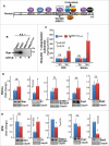
Figure 1.
Figure 1.
Age-dependent deregulation of the ubiquitin-proteasome system (UPS) by Ran-GTP-dependent loss of RBD3 function of Ranbp2. (A) Primary structure of Ranbp2 and partners of domains of Ranbp2. The W2186R mutation in RBD3 of Ranbp2 is shown. LD, leucine-rich domain; RBDn = 1–4, Ran GTPase-binding domains, n = 1–4; ZnFn = 5–8 zinc-finger-rich domain – the number of zinc-fingers vary from 5 to 8 depending on the species (n = 5–8) and they conform to the consensus motif W-XC-X(2,4)-C-X(3)-N-X(6)-C-X (2)-C; KBD, kinesin-1-binding domain; CLD, cyclophilin-like domain; IR, internal repeat; O, overlapping domain between CLD and IR1; CY, cyclophilin domain. (B) GST-fused RDB3 binds Ran GTPase in the presence of the non-hydrolyzable GTP analog, GTP-γ-S, whereas the W2186R mutation in RBD3 abolishes its association with Ran-GTP in GST pull-down assays with retinal extracts (R.E.). (C) Age-dependent accumulation of ubiquitin and ubiquitylated substrates in the RPE and retina. Results are given as means ± s.d.; n = 4. (D, E) The retina (D) and RPE (E) of TgRBD3*-HA::−/− mice share the upregulation of the S1 subunit (also called Psmd1) of the 19S cap of the proteasome. Representative immunoblots are shown below the graphs. Results are given as mean ± s.d.; n = 4. Legend: Exp1, exportin-1/CRM1; S1, S1 subunit of the 19S cap of the proteasome (also called Pmsd1); Ran, Ran GTPase; SUMO-1 RanGAP, SUMOylated Ran GTPase-activating protein (90 kDa); Stat3, signal transducer and activator of transcription 3; m, months, n.s., not significant.
Figure 2.
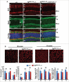
Figure 2.
Confocal microscopy and morphometric analyses of M- and S-cone photoreceptor neurons of 8-week old TgRBD3*-HA::−/− mice. (A) Confocal images of radial sections of the central region of retinae of +/+, TgRBD3*-HA::−/− and TgRBD2/3*-HA::cone-cre::−/− mice immunostained with the pan-cone marker, arrestin (Arr4), and ubc9. +/+ mice present intense and uniform immunostaining of Arr4, whereas such staining is sparser in TgRBD3*-HA::−/− in which cones appear disordered compared with +/+ mice. TgRBD2/3*-HA::cone-cre::−/− mice present much fewer cones than TgRBD3*-HA::−/− mice. The overall distribution of ubc9 in retinal neurons is indistinguishable between genotypes. Note the enrichment of ubc9 in the mitochondrial-rich ellipsoid (E) compartment of rod and cone photoreceptors. Inset in (b) denotes the dash box region of a cone photoreceptor in a-c and the strong labeling of the apical nuclear region (arrow) and ellipsoid (arrowhead). (B) Topographic distribution and length of outer segments of M and S-cone photoreceptors in retinae of +/+ and TgRBD3*-HA::−/− mice. There were no significant differences in the distribution and length of outer segments of M and S-cones in the dorsal (D), central (C) and ventral (V) regions of retinae between genotypes. Images are representative confocal z-stacks images of M and S-cones in the central region of the retina (top panel), whereas quantitative analyses of cones are shown below image panels. Results are given as mean ± s.d.; n = 4. Legend: OS, outer segments; IS, inner segments; ONL, outer nuclear layer (nuclei of photoreceptors); INL, inner nuclear layer (nuclei of inner retinal neurons); E, ellipsoid compartment (of IS); M, myoid compartment (of IS); Syn, synaptiC-terminal; D, dorsal; C, central; V, ventral; ns, non-significant; TgRBD3*-HA, Tg-Ranbp2RBD3*-HA; TgRBD2/3*-HA, Tg-Ranbp2RBD2/3*-HA; −/−, Ranbp2/; cone-cre, HRGP-cre; +/+, wild-type; n.s., not significant. Scale bars: 50 μm (A), 20 μm (B).
Figure 3.
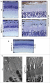
Figure 3.
Light and ultrastructural microscopy of retinas and photoreceptors of +/+ and TgRBD3*-HA::−/− mice. (A, B) Low (A, left panels) and high magnifications (A, right panels) of light microscopy images of methylene blue-stained sections of retinae of 5-month old mice perfused and fixed in 2% paraformaldehyde and 2% glutaraldehyde (A) and 4% glutaraldehyde (B). Note that compared with +/+ mice and when fixed with 2% paraformaldehyde and 2% glutaraldehyde, cone photoreceptors of TgRBD3*-HA::−/− mice present prominent lacunae in OS (arrows) (A), whereas the lacunae in OS are greatly reduced in cones of TgRBD3*-HA::−/− by fixation with 4% glutaraldehyde (B). Black arrowheads point to euchromatic nuclei of cone photoreceptors typically localized in the distal margin of the ONL (A, right panels). (C) Ultrastructural images of photoreceptors of 5-month old mice acquired by transmission electron microscopy. Electrolucent lumina of prominent lacunae in the outer segments (OS) of cone photoreceptors are present only in TgRBD3*-HA::−/− mice. White arrowheads point to euchromatic nuclei of cone photoreceptors with electrolucent chromatin and they contrast with the heterochromatic and electrodense nuclei of rod photoreceptors. Legend: OS, outer segments; IS, inner segments; ONL, outer nuclear layer (nuclei of photoreceptors); INL, inner nuclear layer (nuclei of inner retinal neurons); GC, ganglion neurons; RPE, retinal pigment epithelium; CN, cone nucleus; GA, glutaraldehyde; PA, paraformaldehyde; TgRBD3*-HA, Tg-Ranbp2RBD3*-HA; −/−, Ranbp2−/−; +/+, wild-type. Scale bars: 50 μm (A, left panel; B), 10 μm (A, right panel), 2 μm (C, left panel); 4 μm (C, right panel).
Figure 4.
Figure 4.
Identification of an ubc9 isoform, which selectively localizes to the mitochondrial fraction. (A) Immunoblot of ubc9 of mouse retinal homogenates shows that ubc9 resolves in SDS-PAGE as 2 species of molecular masses of < 25 and >35 kDa. (B) Immunoblot of Ubc9 of subcellular fractions of mouse retinal extracts shows that the low 17 kDa (ubc9) and high molecular ∼36 kDa isoforms (ubc9*) of ubc9 localize to the cytosolic (Cyt) and mitochondrial fractions (Mit), respectively. Gadph and mHsp70 are cytosolic and mitochondrial markers, respectively. (C) Immunoblot of Ubc9 of subcellular fractions of 3T3 fibroblast extracts shows that the ∼17 (ubc9) and ∼36 kDa isoforms (ubc9*) of ubc9 localize to the cytosolic (Cyt) and mitochondrial fractions (Mit), respectively (left panel). The 24 and 34 kDa isoforms of unmodified (Ran) and SUMOylated Ran GTPase (Ran*), respectively, are excluded from the mitochondrial fraction and both are found in the cytosolic fraction (middle panel). Gadph and mHsp70 are cytosolic and mitochondrial markers, respectively (right panel). Legend: Ran, Ran GTPase; Gadph, glyceraldehyde 3-phosphate dehydrogenase; mHsp70, mitochondrial heat shock protein 70.
Figure 5.
Figure 5.
Identification of a Ranbp2 isoform, which selectively associates with mitochondrial markers and localizes to the mitochondrial fraction. (A) Antibodies against Ranbp2 and selective domains thereof. The antibodies, ZnF Ab08 and ZnF Ab09, were raised against the first 5 zinc-finger (ZnF) domains of Ranbp2, KBD Ab41 was raised against the KBD domain of Ranbp2 and mAb414 recognizes undefined epitopes shared by Ranbp2 (Nup358), Nup68 and Nup153. (B) The mAb414 recognizes Ranbp2 (Nup358), Nup68 and Nup153 in retinal homogenates. (C) The ZnF Ab09 recognizes a low molecular mass isoform of Ranbp2 of ∼80 kDa (Ranbp2i80) in co-immunoprecipitates of retinal extracts with the Ranbp2 antibodies, ZnF Ab09 and KBD Ab41, and antibodies against the mitochondrial proteins, mHsp70 and cox11. The large 358 kDa isoform of Ranbp2 (Ranbp2i358) is not co-immunoprecipitated from extracts by the antibodies against mHsp70, cox11and HK1 (lanes 5, 6 and 7). HK1 antibody does not co-immunoprecipitate Ranbp2i80 (lane 7). Ranbp2i358 is co-immunoprecipitated from retinal extracts with the antibodies, mAb414, ZnF Ab09 and KBD Ab41. (D) The Ranbp2 antibody, KBD Ab41, detects only the small Ranbp2i80 in co-immunoprecipitates of retinal extracts with the mHsp70 antibody. (E) Blocking of antibody, ZnF Ab09, with purified recombinant ZnF protein abolishes the detection of Ranbp2i80 co-immunoprecipitated from retinal extracts with mHsp70 antibody. (F) The Ranbp2 antibody, ZnF Ab09, detects only the Ranbp2i80 isoform in purified mitochondria (Mit) immunoprecipitated with antibodies against mHsp70 and cox11. (G) mHsp70 is co-immunoprecipitated from retinal extracts by the Ranbp2 antibodies, ZnF Ab09 and KBD Ab41. IgG (rabbit) is a control antibody in C-G. The murine origin of the monoclonal antibodies, mAb414, mHsp70 and HK1, leads to weak signals of IgGH in panels, (C)and F, owing to poor cross-reactivity with the goat anti-rabbit enzyme (HRP)-conjugated secondary antibody. Legends: R.E., retinal extracts; IgGH, IgG heavy-chain; IgGL, IgG light-chain; IB, immunoblot; IP, immunoprecipitation; HK1, hexokinase-1; mHsp70, mitochondrial heat shock protein 70.
Figure 6.
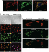
Figure 6.
Liposome-mediated delivery of anti-KBD antibody of Ranbp2 into 661W neurons leads to mitochondrial fragmentation, delocalization of RanBP2 from mitochondria and neuronal atrophy. (A) Ranbp2 colocalizes extensively with mHsp70-immunostained worm-like mitochondria in 661W neurons. Ranbp2 was detected with ZnF Ab09. (B) Ranbp2 colocalizes extensively with mHsp70-immunostained worm-like mitochondria in liposome mock-treated cells and cells present healthy morphology (a-d). Liposome-mediated delivery of KBD Ab41 into 661W neurons (e-h) causes strong neural atrophy (e), mitochondrial fragmentation (g) and the delocalization of Ranbp2 from the mitochondria (h) without affecting the localization of Ranbp2 at the nuclear rim (f). Internalized KBD Ab41 was detected with Alexa-conjugated goat anti-rabbit antibody (f). (C) High-magnification images of liposome mock and KBD Ab41-treated 661W neurons immunostained for Ranbp2 and mHsp70 as shown in image panels of B. Legends: KBD Ab41 Transf., cells with liposome-mediated delivery of Ranbp2 antibody, KBD Ab41; Mock Transf., liposome mock-treated cells. DIC, differential interference contrast (Nomarski optics). Scale bars: 10 μm (A), 30 μm (B), 20 μm (C).

Similar articles

Cited by

References

    1. Delphin C, Guan T, Melchior F, Gerace L. RanGTP targets p97 to RanBP2, a filamentous protein localized at the cytoplasmic periphery of the nuclear pore complex. Mol Biol Cell. 1997;8(12):2379-90. doi:10.1091/mbc.8.12.2379. PMID:9398662 - DOI - PMC - PubMed
    1. Vetter IR, Nowak C, Nishimoto T, Kuhlmann J, Wittinghofer A. Structure of a Ran-binding domain complexed with Ran bound to a GTP analogue: implications for nuclear transport. Nature. 1999;398(6722):39-46. doi:10.1038/17969. PMID:10078529 - DOI - PubMed
    1. Hamada M, Haeger A, Jeganathan KB, van Ree JH, Malureanu L, Walde S, Joseph J, Kehlenbach RH, van Deursen JM. Ran-dependent docking of importin-beta to RanBP2/Nup358 filaments is essential for protein import and cell viability. J Cell Biol. 2011;194(4):597-612. doi:10.1083/jcb.201102018. PMID:21859863 - DOI - PMC - PubMed
    1. Ritterhoff T, Das H, Hofhaus G, Schroder RR, Flotho A, Melchior F. The RanBP2/RanGAP1*SUMO1/Ubc9 SUMO E3 ligase is a disassembly machine for Crm1-dependent nuclear export complexes. Nat Commun. 2016;7:11482. doi:10.1038/ncomms11482. PMID:27160050 - DOI - PMC - PubMed
    1. Singh BB, Patel HH, Roepman R, Schick D, Ferreira PA. The zinc finger cluster domain of RanBP2 is a specific docking site for the nuclear export factor, exportin-1. J Biol Chem. 1999;274(52):37370-8. doi:10.1074/jbc.274.52.37370. PMID:10601307 - DOI - PubMed

Publication types

MeSH terms