. 2018 Jan;42(1):28-38.
doi: 10.1097/PAS.0000000000000938.

Recurrent BRAF Gene Fusions in a Subset of Pediatric Spindle Cell Sarcomas: Expanding the Genetic Spectrum of Tumors With Overlapping Features With Infantile Fibrosarcoma

Affiliations

Recurrent BRAF Gene Fusions in a Subset of Pediatric Spindle Cell Sarcomas: Expanding the Genetic Spectrum of Tumors With Overlapping Features With Infantile Fibrosarcoma

Yu-Chien Kao et al. Am J Surg Pathol. 2018 Jan.

Abstract

Infantile fibrosarcomas (IFS) represent a distinct group of soft tissue tumors occurring in patients under 2 years of age and most commonly involving the extremities. Most IFS show recurrent ETV6-NTRK3 gene fusions, sensitivity to chemotherapy, and an overall favorable clinical outcome. However, outside these well-defined pathologic features, no studies have investigated IFS lacking ETV6-NTRK3 fusions, or tumors with the morphology resembling IFS in older children. This study was triggered by the identification of a novel SEPT7-BRAF fusion in an unclassified retroperitoneal spindle cell sarcoma in a 16-year-old female by targeted RNA sequencing. Fluorescence in situ hybridization screening of 9 additional tumors with similar phenotype and lacking ETV6-NTRK3 identified 4 additional cases with BRAF gene rearrangements in the pelvic cavity (n=2), paraspinal region (n=1), and thigh (n=1) of young children (0 to 3 y old). Histologically, 4 cases including the index case shared a fascicular growth of packed monomorphic spindle cells, with uniform nuclei and fine chromatin, and a dilated branching vasculature; while the remaining case was composed of compact cellular sheets of short spindle to ovoid cells. In addition, a minor small blue round cell component was present in 1 case. Mitotic activity ranged from 1 to 9/10 high power fields. Immunohistochemical stains were nonspecific, with only focal smooth muscle actin staining demonstrated in 3 cases tested. Of the remaining 5 BRAF negative cases, further RNA sequencing identified 1 case with EML4-NTRK3 in an 1-year-old boy with a foot IFS, and a second case with TPM3-NTRK1 fusion in a 7-week-old infant with a retroperitoneal lesion. Our findings of recurrent BRAF gene rearrangements in tumors showing morphologic overlap with IFS expand the genetic spectrum of fusion-positive spindle cell sarcomas, to include unusual presentations, such as older children and adolescents and predilection for axial location, thereby opening new opportunities for kinase-targeted therapeutic intervention.


Conflict of interest statement

Conflicts of interest: none

Figures

Figure 1
Figure 1
Histologic features of the index case of a retroperitoneal tumor in a 16 year-old female. The tumor is composed of uniform spindle cells associated with scattered dilated branching vessels (A) and arranged in intersecting fascicles (B). The tumor cells have elongated nuclei, mild atypia, a mitotic count up to 6/10 high power fields, and accompanied by a variable lymphocytic infiltrate (C). Immunohistochemical stains showed focal smooth muscle actin staining (D).
Figure 2
Figure 2
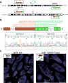
A SEPT7-BRAF gene fusion was identified in the index case by targeted RNA sequencing. A pericentric inversion of chromosome 7 resulted in the fusion of SEPT7 (7p14.2) and BRAF (7q34) (A). RNA sequencing fusion junction reads and subsequent confirmatory RT-PCR showed SEPT7 exon 11 fused in-frame to BRAF exon 11 (B). FISH further confirmed BRAF gene rearrangement with break-away centromeric (red) and telomeric (green) signals (C, left) as well as SEPT7-BRAF fusion with come-together SEPT7 (red) and BRAF (green) signals (C, right).
Figure 3
Figure 3
Histologic features of 4 additional pediatric spindle cell sarcomas resembling IFS with BRAF rearrangements. Both case 2 (A-C) and case 3 (D-F) occurred in the pelvis, showing a monomorphic spindle cell phenotype arranged in fascicles (B,E) and with scattered dilated vessels (A,D). The tumor cells show fine chromatin pattern (C,E,F). Focal areas of round cell component were also seen in case 3 (F). Case 4 (G), a paraspinal mass, was composed of short spindle to ovoid cells, arranged in dense sheets and focal fascicular growth. Case 5, a congenital thigh mass, showed long spindle cell fascicles with focal myxoid stroma (H) and skeletal muscle infiltration (I).
Figure 4
Figure 4
EML4-NTRK3 fusion in an IFS involving the foot of a 21 month-old boy (case #6). Targeted RNA sequencing showed fusion of EML4 exon 2 to NTRK3 exon 14, resulting from a t(2;15) translocation (A,B). The chimeric protein contains tyrosine kinase domain from NTRK3 but no known functional domain from EML4 (B). The downstream exons after the breakpoint of NTRK3 (exon 14) showed up-regulated mRNA level in contrast to the 5’ NTRK3 exons and other samples in the same dataset (B, orange dots). FISH confirmed rearrangement of both EML4 and NTRK3 genes, with break-apart of centromeric (red) and telomeric (green) signals (C).
Figure 5
Figure 5
TPM3-NTRK1 fusion in a 7 week-old infant retroperitoneal IFS (case #7). Whole transcriptome RNA sequencing identified a 2.7 Mb inversion-fusion at 1q21.3 locus, leading to a TPM3-NTRK1 candidate gene fusion (A). The RT-PCR confirmed an in-frame fusion of TPM3 exon 6 to NTRK1 exon 9 (B). FISH also confirmed NTRK1 rearrangement (split of green and yellow signals) and fusion of TPM3 (red) with NTRK1 (green, telomeric/3’ of NTRK1) (C). NTRK1 mRNA expression was up-regulated compared to other samples in the same platform (D, left). Exonic expression levels showed high expression of NTRK1 exons downstream to the break in exon 9; the fusion retaining the entire tyrosine kinase domain in the chimeric protein (D, right).
Figure 6
Figure 6
Histologic features of IFS with EML4-NTRK3 (case #6; A,B) and TPM3-NTRK1 fusions (case #7; C,D). Case #6 showed primitive round, ovoid to short spindle cells with scattered lymphocytic infiltration (A). The tumor cells had monomorphic nuclei, fine chromatin, and scant cytoplasm (B). Case #7 (primary resection) showed long intersecting fascicles accompanied by cleft-like or compressed vasculature (C, D). The tumor cells had eosinophilic cytoplasm and generally fine chromatin, with scattered atypical hyperchromatic cells (D).

References

    1. Chung EB, Enzinger FM. Infantile fibrosarcoma. Cancer. 1976;38:729–739. - PubMed
    1. Orbach D, Rey A, Cecchetto G, et al. Infantile fibrosarcoma: management based on the European experience. J Clin Oncol. 2010;28:318–323. - PubMed
    1. Soule EH, Pritchard DJ. Fibrosarcoma in infants and children: a review of 110 cases. Cancer. 1977;40:1711–1721. - PubMed
    1. Nagasubramanian R, Wei J, Gordon P, et al. Infantile Fibrosarcoma With NTRK3-ETV6 Fusion Successfully Treated With the Tropomyosin-Related Kinase Inhibitor LOXO-101. Pediatr Blood Cancer. 2016;63:1468–1470. - PMC - PubMed
    1. Knezevich SR, McFadden DE, Tao W, et al. A novel ETV6-NTRK3 gene fusion in congenital fibrosarcoma. Nat Genet. 1998;18:184–187. - PubMed

MeSH terms