. 2017 Sep 6;12(9):e0183214.
doi: 10.1371/journal.pone.0183214. eCollection 2017.

Functional characterization of T-cells from palatine tonsils in patients with chronic tonsillitis

Affiliations

Functional characterization of T-cells from palatine tonsils in patients with chronic tonsillitis

Katharina Geißler et al. PLoS One. .

Abstract

The palatine tonsils, localized in the oropharynx, are easily accessible secondary lymphoid tissue in humans. Inflammation of the palatine tonsils, local and chronic in case of chronic tonsillitis (CT) or acute in the presence of a peritonsillar abscess (PTA), ranks among the most common diseases in otolaryngology. However, the functionality of tonsillar immune cells, notably T-cells, in the context of these immune pathologies is poorly understood. We have examined the functional status of human tonsillar T-cells in CT and compared it to the acute inflammatory setting of a PTA. Patients presenting with CT (n = 10) or unilateral PTA (n = 7) underwent bilateral tonsillectomy and a subgroup of 8 patients underwent additional blood sampling. T-cells were purified via automated magnetic selection and subjected to flow cytometry-based immunophenotyping. In addition, the response to T-cell receptor (TCR) stimulation was assessed at the level of proximal signaling, activation marker expression and proliferation. We observed no difference between the percentage of T helper (CD4(+)) cells from tonsil tissue in CT and PTA, but observed a trend towards a higher percentage of T helper cells in the blood of patients with PTA versus CT, probably reflecting an acute, systemic bacterial infection in the former cohort. Tonsils from CT harbored more PD-1(+) CD4(+) T-cells, pointing to T-cell exhaustion due to chronic infection. This notion was supported by functional studies that showed a tendency to weaker TCR responses of tonsillar T-cells from CT. Intriguingly, tonsillar T-cells recurrently featured a dampened response to T-cell receptor stimulation at the level of receptor proximal signaling steps compared to peripheral T-cells. In sum, our study documents distinct differences in tonsillar T-cell class distribution and function between the various pathological conditions. Our observations are consistent with the concept that tonsillar T-cells react to infections by eliciting specific immunological responses in chronic versus acute settings of inflammation.


Conflict of interest statement

Competing Interests: The authors have declared that no competing interests exist.

Figures

Fig 1
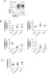
Fig 1. Tonsillar CD4/CD8 T-cell counts are not affected by the particular inflammation scenarios.
(A) Purity of tonsillar T-cells isolated by positive magnetic selection as determined by surface CD3 staining. A representative flow cytometry histogram depicting CD3 staining of purified CD4(+)CD8(+) T-cells (black profile) and the residual negative cellular fraction (grey profile) is shown. Insert describes the purity in % CD3 positive of total tonsillar cells. (B) The surface expression of CD4 and CD8 on purified tonsillar and peripheral T-cells was assessed by flow cytometry and plotted as % CD4(+) of total T-cells and CD4(+)/CD8(+) ratio. For statistical analysis, see S1 Table. (C) CD4(+)/CD8(+) T-cell numbers from tonsils with abscess and their paired healthy counterparts. Data are plotted as CD4(+)/CD8(+) ratio. Data are presented as mean + SEM. CT = chronic tonsillitis; PTA = peritonsillar abscess; HY = tonsillar hyperplasia; ton = tonsil; blo = blood; abs = abscess; hea = healthy.
Fig 2
Fig 2. Tonsillar T-cell immune phenotype.
CD4(+) plus CD8(+) T-cells were purified from tonsillar samples followed by fluorescence assisted immune staining of CD45RA and CD62L for the determination of naïve/effector/memory T-cells. All shown measurements were performed on the mixed CD4(+)CD8(+) preparations. See text for further details. (A) Representative histogram of a double staining for CD45RA and CD62L. (B) Quantification of all single and double positive CD45RA/CD62L populations for the various tonsillar disease groups. (C) Flow cytometric determination of CD45R0 expression on tonsillar T-cells. CT = chronic tonsillitis; PTA = peritonsillar abscess; HY = tonsillar hyperplasia; ton = tonsil; abs = abscess.
Fig 3
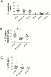
Fig 3. Inhibitory and suppressed T-cell populations in tonsils.
CD4(+) and CD8(+) T-cells purified from tonsillar samples were subjected to fluorescence assisted immune phenotyping for the determination of inhibitory T-cell subgroups. All shown measurements were carried out on the mixed CD4(+)CD8(+) preparations. (A) The incidence of regulatory T-cells (Tregs) was determined by triple CD4/CD25/FoxP3 staining and plotted as percentage of Tregs in all T-cells both for tonsillar and blood samples. For statistical analysis, see suppl.S1 Table. (B) The fraction of PD-1 expressing T-cells was determined by flow cytometric determination of PD-1 cell surface staining and plotted as percentage of PD-1(+) cells in CD4(+) lymphocytes (CT-ton vs. PTA-abs.ton p = 0.026). (C) The surface expression of CTLA-4 on tonsillar T-cells was determined by flow cytometry and plotted as percentage of CTLA4(+) in all T-cells. * indicates comparisons that met an unadjusted p≤0.05 (explorative significance level). CT = chronic tonsillitis; PTA = peritonsillar abscess; HY = tonsillar hyperplasia; ton = tonsil; abs = abscess.
Fig 4
Fig 4. Tonsillar T-cells from chronic tonsillitis are basally activated.
CD4(+)/CD8(+) T-cells were isolated from tonsils of the different disease scenarios and their activation status was determined via flow cytometry analysis of the mixed CD4(+)CD8(+) preparations. (A) CD69, CD25 and CD154 activation marker surface expression determined by flow cytometry and plotted as percentage of all T-cells. (B) Double immune-staining for PD-1 and CD69 in all T-cells. Only statistically significant groups are labelled (* p<0.05). CT = chronic tonsillitis; PTA = peritonsillar abscess; HY = tonsillar hyperplasia; ton = tonsil; abs = abscess.
Fig 5
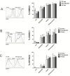
Fig 5. Tonsillar T-cells are not compromised in activation marker upregulation in response to TCR activation.
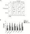
CD4/CD8 T-cells isolated from tonsils were challenged with the indicated formulations of anti CD3 and/or CD28 Abs or with a mix of phorbol ester and Ionomycine (TPA/Iono) and subjected to flow cytometric determination of activation marker CD69 (A), CD25 (B) and CD154 (C) expression, always plotted as percentage of all T-cells. All measurements were carried out on the mixed CD4(+)CD8(+) preparations. Representative fluorescence profiles for stimulated (black line) versus non-stimulated samples (grey curves) are shown on the left side of each panel. Beads: anti-CD3 and anti-CD28 Abs immobilized on beads. CT = chronic tonsillitis; PTA = peritonsillar abscess; HY = tonsillar hyperplasia; ton = tonsil; abs = abscess.
Fig 6
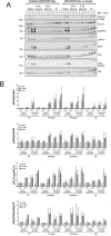
Fig 6. Analysis of TCR-proximal signal transduction in tonsillar T-cells.
(A) CD4/CD8 T-cells were isolated from tonsils and resuspended in FCS-free medium. The total CD4(+)CD8(+) T-cell preparations were challenged with soluble or immobilized anti CD3/CD28 Abs to activate the TCR. Cells were lysed and cellular extracts were processed for Western Blot detection of total and phosphorylated versions of PLCγ, ZAP70, Erk and Akt. (B) Bands were quantified by densitometry and the ratio of phosphorylated to total protein was plotted as as fold activation of unstimulated samples (0 min). The quantification includes all measured tonsil samples. Note that the values for PTA-abs/blo and PTA-heal/blo represent perforce the same data and are plotted twice for illustrative reasons. Data are presented as mean + SEM. pPLCγ, pErk, pAkt and pZAP-70 mark the phosphorylated versions of the respective proteins. CT = chronic tonsillitis; PTA = peritonsillar abscess; HY = tonsillar hyperplasia; ton = tonsil; blo = blood; abs = abscess; hea = healthy.
Fig 7
Fig 7. Tonsillar T-cells expand and proliferate in response to TCR activation.
(A) CD4/CD8 T-cells isolated from tonsils were loaded with the vital dye CFSE and challenged with soluble or immobilized anti-CD3/CD28 Abs to activate the TCR. Cell proliferation of the mixed CD4(+)CD8(+) T-cell preparations was scored as a reduction in CFSE fluorescence as T-cells divide and segregate the dye to daughter cells. Shown here are representative profiles for one sample of each disease group. Insert numbers represent the percentage of all T-cells that underwent at least one cell division. (B) Quantification of cell proliferation for all tonsil samples performed with FlowJo software (TreeStar Inc., Ashland, USA). For statistical evaluation, see S1 Table. CT = chronic tonsillitis; PTA = peritonsillar abscess; HY = tonsillar hyperplasia; ton = tonsil; blo = blood; abs = abscess; hea = healthy.

References

    1. Nave H, Gebert A, Pabst R. Morphology and immunology of the human palatine tonsil. Anat Embryol (Berl). 2001;204(5):367–73. - PubMed
    1. Olofsson K, Hellstrom S, Hammarstrom ML. The surface epithelium of recurrent infected palatine tonsils is rich in gammadelta T cells. Clin Exp Immunol. 1998;111(1):36–47. doi: 10.1046/j.1365-2249.1998.00446.x - DOI - PMC - PubMed
    1. Palomares O, Ruckert B, Jartti T, Kucuksezer UC, Puhakka T, Gomez E, et al. Induction and maintenance of allergen-specific FOXP3+ Treg cells in human tonsils as potential first-line organs of oral tolerance. J Allergy Clin Immunol. 2012;129(2):510–20, 20.e1-9. doi: 10.1016/j.jaci.2011.09.031 - DOI - PubMed
    1. Burton MJ, Glasziou PP. Tonsillectomy or adeno-tonsillectomy versus non-surgical treatment for chronic/recurrent acute tonsillitis. Cochrane Database Syst Rev. 2009(1):CD001802 doi: 10.1002/14651858.CD001802.pub2 - DOI - PubMed
    1. Paradise JL, Bluestone CD, Bachman RZ, Colborn DK, Bernard BS, Taylor FH, et al. Efficacy of tonsillectomy for recurrent throat infection in severely affected children. Results of parallel randomized and nonrandomized clinical trials. N Engl J Med. 1984;310(11):674–83. doi: 10.1056/NEJM198403153101102 - DOI - PubMed

MeSH terms