. 2017 Sep 5;26(3):568-575.e3.
doi: 10.1016/j.cmet.2017.08.013.

Persistence of Pancreatic Insulin mRNA Expression and Proinsulin Protein in Type 1 Diabetes Pancreata

Affiliations

Persistence of Pancreatic Insulin mRNA Expression and Proinsulin Protein in Type 1 Diabetes Pancreata

Clive Wasserfall et al. Cell Metab. .

Abstract

The canonical notion that type 1 diabetes (T1D) results following a complete destruction of β cells has recently been questioned as small amounts of C-peptide are detectable in patients with long-standing disease. We analyzed protein and gene expression levels for proinsulin, insulin, C-peptide, and islet amyloid polypeptide within pancreatic tissues from T1D, autoantibody positive (Ab+), and control organs. Insulin and C-peptide levels were low to undetectable in extracts from the T1D cohort; however, proinsulin and INS mRNA were detected in the majority of T1D pancreata. Interestingly, heterogeneous nuclear RNA (hnRNA) for insulin and INS-IGF2, both originating from the INS promoter, were essentially undetectable in T1D pancreata, arguing for a silent INS promoter. Expression of PCSK1, a convertase responsible for proinsulin processing, was reduced in T1D pancreata, supportive of persistent proinsulin. These data implicate the existence of β cells enriched for inefficient insulin/C-peptide production in T1D patients, potentially less susceptible to autoimmune destruction.

Keywords: PCSK1; PCSK2; insulin mRNA; insulin promoter; insulin-positive single cells; pancreas; proconvertases; proinsulin; type 1 diabetes.


Figures

Figure 1
Figure 1. Proinsulin, insulin, and glucagon expression in type 1 diabetes (T1D)
(A–E) Composite images of pancreas sections stained for proinsulin (green), insulin (red), glucagon (yellow), and DAPI (nuclei, blue) are shown for sections containing islets from (A) a control 24-year old male Caucasian (nPOD 6131), (B–C) a 12-year old male African American with T1D for 1 year (nPOD 6052), (D) a 23-year old female Caucasian with T1D for 7 years (nPOD 6070), and (E) a section containing pancreas exocrine tissue from a 12.5 year old female Caucasian with T1D for 2 years (nPOD 6371). (F–T) The individual channels for (F–J) proinsulin (green), (K–O) insulin (red), and (P–T) glucagon (yellow) are also shown. Insets display: (A) proinsulin and insulin positive cells in the exocrine pancreas, (C) an insulin negative, proinsulin negative, glucagon positive islet and (E) proinsulin, insulin and glucagon positive cells in the exocrine pancreas. Scale bars represent 50 μm. See also Figure S1
Figure 2
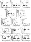
Figure 2. Total protein extracts and gene expression from human pancreas sections
(A–H) ELISAs of acid-ethanol extracts from human pancreas tissue sections for (A) Insulin (μInternational Units (μIU)), (B) Proinsulin, (C) C-Peptide, (D) Islet Amyloid Polypeptide, and (E) Glucagon (values were normalized against total protein). ND = not detected/examined are noted on each graph. Data are presented as median with interquartile range. (F–H) Correlation between extracted proinsulin and extracted insulin in (F) Controls, (G) autoantibody positive (Ab+), and (H) T1D is shown. (H) For T1D subjects, correlation analysis is reported for all data as well as a separate analysis of the five subjects with insulin detected in the normal range (Detectable INS Only) with the latter represented by the trend line. (I–N) Real time qPCR Cq values for control and T1D pancreata for (I) INS, (J) unspliced INS heterogeneous nuclear RNA (hnRNA), (K) INS-IGF2 readthrough mRNA, (L) IAPP, (M) GCG, and (N) SST expression were compared. For INS hnRNA, INS-IGF2, and IAPP, statistical analyses were not performed due to the number of T1D samples with undetectable RNA (ND = not detected/examined). p-values are indicated on the figure.
Figure 3
Figure 3. Evaluation of insulin/glucagon IHC, INS ISH, prohormone convertase and protease gene expression from human pancreas
(A–E) Insulin (red) and glucagon (blue) protein was detected by IHC, and (F–J) insulin mRNA (pink) was detected by ISH in pancreas sections from donors without diabetes (A,F; Control; nPOD 6172) and T1D pancreata with 7 (B,C,G,H; nPOD 6070) and 35 year duration (D,E,I,J; nPOD 6031). Insets in (D) and (E) illustrate glucagon only positive islets. Red arrows indicate single insulin positive cells located in the exocrine tissue and peri-ductal. Scale bars represent 50μm. (K–O) For control (n=5) and T1D subjects (n=11), IHC analysis of glucagon and insulin within pancreas sections from control donors as well as short (0–7 years; n=5) or long (8–35 years; n=6) duration T1D were analyzed for (K) insulin positive cells per islet; cell counts within the (L–M) islets and (N–O) exocrine tissue. (P–R) qPCR for control versus T1D pancreata (P) PCSK1, (Q) PCSK2, and (R) CPE. p-values indicated on the figure.

References

    1. Alarcon C, Boland BB, Uchizono Y, Moore PC, Peterson B, Rajan S, Rhodes OS, Noske AB, Haataja L, Arvan P, et al. Pancreatic beta-Cell Adaptive Plasticity in Obesity Increases Insulin Production but Adversely Affects Secretory Function. Diabetes. 2016;65:438–450. - PMC - PubMed
    1. Andersson AK, Sandler S. Melatonin protects against streptozotocin, but not interleukin-1beta-induced damage of rodent pancreatic beta-cells. Journal of pineal research. 2001;30:157–165. - PubMed
    1. Atkinson MA, Eisenbarth GS, Michels AW. Type 1 diabetes. Lancet. 2014;383:69–82. - PMC - PubMed
    1. Campbell-Thompson M. Organ donor specimens: What can they tell us about type 1 diabetes? Pediatr Diabetes. 2015;16:320–330. - PMC - PubMed
    1. Campbell-Thompson M, Fu A, Kaddis JS, Wasserfall C, Schatz DA, Pugliese A, Atkinson MA. Insulitis and beta-Cell Mass in the Natural History of Type 1 Diabetes. Diabetes. 2016;65:719–731. - PMC - PubMed