. 2017 Oct 20;292(42):17449-17460.
doi: 10.1074/jbc.M117.807396. Epub 2017 Sep 6.

Structural basis for pH-insensitive inhibition of immunoglobulin G recycling by an anti-neonatal Fc receptor antibody

Affiliations

Structural basis for pH-insensitive inhibition of immunoglobulin G recycling by an anti-neonatal Fc receptor antibody

Jon A Kenniston et al. J Biol Chem. .

Abstract

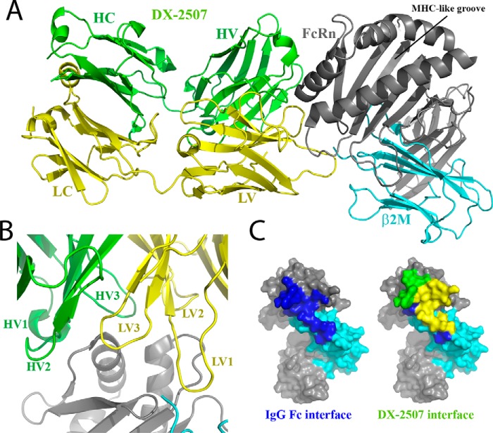
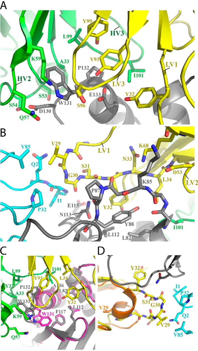
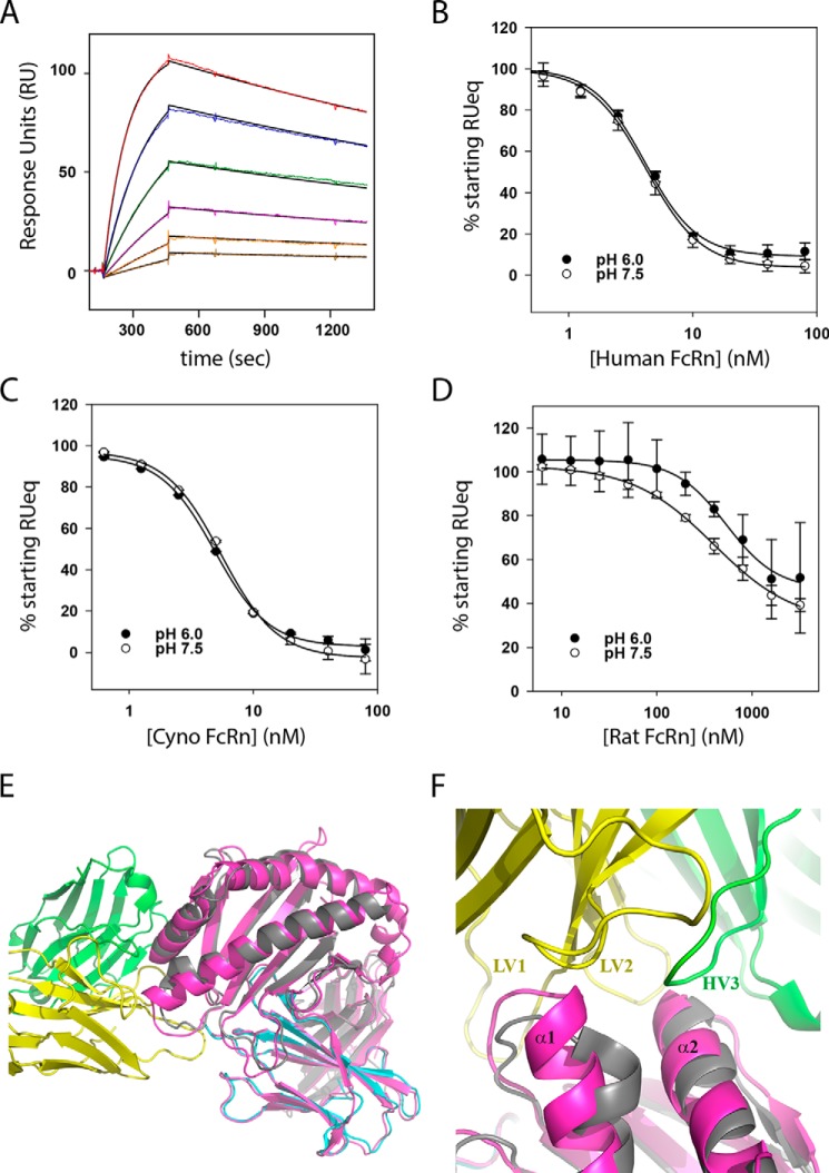
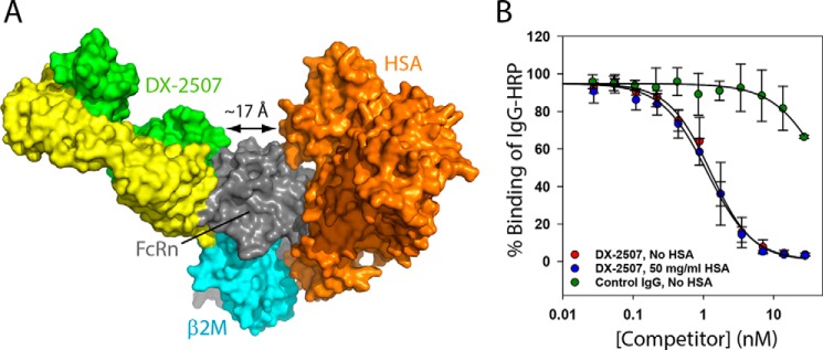
The neonatal Fc receptor FcRn plays a critical role in the trafficking of IgGs across tissue barriers and in retaining high circulating concentrations of both IgG and albumin. Although generally beneficial from an immunological perspective in maintaining IgG populations, FcRn can contribute to the pathogenesis of autoimmune disorders when an abnormal immune response targets normal biological components. We previously described a monoclonal antibody (DX-2507) that binds to FcRn with high affinity at both neutral and acidic pH, prevents the simultaneous binding of IgG, and reduces circulating IgG levels in preclinical animal models. Here, we report a 2.5 Å resolution X-ray crystal structure of an FcRn-DX-2507 Fab complex, revealing a nearly complete overlap of the IgG-Fc binding site in FcRn by complementarity-determining regions in DX-2507. This overlap explains how DX-2507 blocks IgG binding to FcRn and thereby shortens IgG half-life by preventing IgGs from recycling back into circulation. Moreover, the complex structure explains how the DX-2507 interaction is pH-insensitive unlike normal Fc interactions and how serum albumin levels are unaffected by DX-2507 binding. These structural studies could inform antibody-based therapeutic approaches for limiting the effects of IgG-mediated autoimmune disease.

Keywords: Fc receptor; antibody; autoimmune disease; immunoglobulin G (IgG); inhibitor; receptor recycling.


Conflict of interest statement

This investigation was supported by Dyax Corp., now a wholly owned subsidiary of Shire Pharmaceuticals. J. A. K., G. P. C., J. C., K. J. K., A. P. L., S. R. C., C. T., B. A. A., D. J. S., and A. E. N. were employees of Dyax at the time of the study. J. A. K. and D. J. S. are currently Shire employees

Figures

Figure 1.
Figure 1.
Equivalent targeting of human FcRn by the DX-2507 IgG and Fab constructs. A, a representative SPR sensorgram of human sFcRn injected at multiple concentrations over surface-immobilized DX-2507 IgG. Color lines show the measured RU value during the association and dissociation phases, whereas the solid black line is the best fit to a 1:1 Langmuir model. Average fits are presented in Table 1. B, SPR sensorgram as in A, but the biosensor surface was coated with DX-2507 Fab. C, flow cytometry experiments show that increasing concentrations of DX-2507 IgG (blue circles) and Fab (red circles) can compete with fluorescent IgG-AF488 binding to HEK293T cells expressing human FcRn (expressed as a shift in the mean FITC peak), in contrast to a control IgG (DX-2300; green triangles) that only targets FcRn through the canonical Fc interaction. In the absence of expressed FcRn, there is a low IgG-AF488 signal that is not competed by DX-2507. Error bars, S.D. of replicates (n ≥ 2); solid lines, fit to an IC50 equation.
Figure 2.
Figure 2.
Crystal structures of apo and complexed DX-2507 Fab. A, overlay of the apo-DX-2507 Fab structure (PDB code 5WHJ) with the Fab domain that is bound to FcRn (PDB code 5WHK, RMSD = 0.733 Å). The Fab light chain is shown in brown and yellow ribbon for apo and complexed Fab, respectively, and the heavy chain is shown in pale green and green ribbon for apo and complexed Fab, respectively. Certain CDR loops (e.g. HV2) highlight the region of the DX-2507 Fab that interacts with FcRn. Extension of LV1 from a helical conformation to an extended loop is the most apparent difference, although some minor changes in LV3 and HV2 are evident. B, temperature factor analysis reveals that the FcRn interface residues are generally more ordered in the complex than in the Fab crystallized alone. The dashed circle region highlights the central region of the Fab that interacts with FcRn.
Figure 3.
Figure 3.
X-ray crystal structure of the DX-2507 Fab bound to human FcRn. A, ribbon model of the DX-2507–FcRn complex (PDB code 5WHK). The FcRn heavy chain is colored in gray, and the β2M subunit is shown in cyan. The DX-2507 heavy chain is colored green, and the light chain is yellow. B, close-up view of the DX-2507 CDRs, showing all of them in close proximity to FcRn. C, FcRn is shown as a surface schematic with coloring as in A. Left, the canonical IgG–Fc interface residues are highlighted in blue, and (right) superimposed with residues interacting with the DX-2507 heavy (green) and light (yellow) chains. The DX-2507 epitope covers most of the IgG–Fc binding region of FcRn.
Figure 4.
Figure 4.
A detailed look at the DX-2507–FcRn complex paratope–epitope interface. A, the central interaction between DX-2507 HV2, HV3, LV1, and LV3 and the FcRn stretch of residues 130DWPE133 drives complex formation. The color scheme is as in Fig. 3. B, DX-2507 interacts with a second major linear stretch of FcRn residues Ala81–Tyr88. C, a view of the extensive hydrophobic interactions that define the center of the complex interface. In addition, it is notable that Trp131 is flipped toward the DX-2507 paratope relative to all other reported human FcRn structures, including the Fc-complexed FcRn structure aligned here (magenta; PDB code 4N0U). D, DX-2507 LV1 unfurls from an α-helical conformation in the apo-Fab structure (orange) to an extended loop, as the complexed Fab (yellow) now contacts residues in the β2M subunit of FcRn (cyan).
Figure 5.
Figure 5.
DX-2507 potently binds cyno FcRn but not rat FcRn. A, an example SPR sensorgram of soluble cyno FcRn interacting with surface-immobilized DX-2507 Fab. Color lines show the experimental RU value for the association and dissociation phases, with the solid black line representing the best fit to a 1:1 Langmuir model. B, a competition SPR experiment probes the ability of added FcRn to compete away binding of DX-2507 Fab analyte to surface-immobilized human FcRn. Error bars, S.D. of replicates (n ≥ 2); solid lines, fit to an IC50 equation = 4.1 ± 0.3 nm (pH 6.0) and 4.2 ± 0.2 nm (pH 7.5). C, competition SPR as in B but with increasing concentrations of cyno FcRn as the competitor. Fitted IC50 = 4.9 ± 0.2 nm (pH 6.0) and 5.5 ± 0.2 nm (pH 7.5). D, rat FcRn competition SPR demonstrates a significant decrease in affinity for this species, with resulting IC50 fits = 535 ± 60 nm (pH 6.0) and 397 ± 33 nm (pH 7.5). E, alignment of the DX-2507–shFcRn complex structure (colored as in Fig. 3) with rat FcRn (colored magenta; PDB code 3FRU) shows generally good agreement between the structures (RMSD = 1.07 Å). F, close-up of the alignment of rat and human FcRn at the DX-2507 interface reveals a small but significant extension of α-helix 1 in the rat FcRn that would protrude into DX-2507 LV1 and LV2, probably explaining why this interaction has reduced affinity relative to human FcRn.
Figure 6.
Figure 6.
Binding of DX-2507 to FcRn is independent of HSA binding. A, structural model of the simultaneous binding of DX-2507 and HSA (orange) based on the structural alignment of FcRn from the complex structure in this study (PDB code 5WHK) and FcRn in complex with HSA (PDB code 4K71). HSA and DX-2507 Fab target distinct regions of FcRn. B, the addition of 50 mg/ml HSA (at the high end of the normal physiological concentration in blood) does not influence the ability of DX-2507 to compete away IgG-HRP binding to microplate immobilized FcRn, as exhibited in the competition ELISA experiment shown here. At pH 6.0, a control IgG (DX-2300) competes only at high IgG (Fc) concentrations relative to DX-2507. Error bars, S.D. of replicates (n ≥ 2); solid lines, fit to an IC50 equation.

References

    1. Sesarman A., Vidarsson G., and Sitaru C. (2010) The neonatal Fc receptor as therapeutic target in IgG-mediated autoimmune diseases. Cell Mol. Life Sci. 67, 2533–2550 - PMC - PubMed
    1. Pyzik M., Rath T., Lencer W. I., Baker K., and Blumberg R. S. (2015) FcRn: the architect behind the immune and nonimmune functions of IgG and albumin. J. Immunol. 194, 4595–4603 - PMC - PubMed
    1. Nixon A. E., Chen J., Sexton D. J., Muruganandam A., Bitonti A. J., Dumont J., Viswanathan M., Martik D., Wassaf D., Mezo A., Wood C. R., Biedenkapp J. C., and TenHoor C. (2015) Fully human monoclonal antibody inhibitors of the neonatal fc receptor reduce circulating IgG in non-human primates. Front. Immunol. 6, 176. - PMC - PubMed
    1. Simister N. E., and Mostov K. E. (1989) An Fc receptor structurally related to MHC class I antigens. Nature 337, 184–187 - PubMed
    1. Martin W. L., West A. P. Jr, Gan L., and Bjorkman P. J. (2001) Crystal structure at 2.8 Å of an FcRn/heterodimeric Fc complex: mechanism of pH-dependent binding. Mol. Cell 7, 867–877 - PubMed

Substances

LinkOut - more resources