

Nonlinear temporal dynamics of cerebral small vessel disease: The RUN DMC study
- PMID: 28878046
- PMCID: PMC5634663
- DOI: 10.1212/WNL.0000000000004490
Nonlinear temporal dynamics of cerebral small vessel disease: The RUN DMC study
Abstract
Objective: To investigate the temporal dynamics of cerebral small vessel disease (SVD) by 3 consecutive assessments over a period of 9 years, distinguishing progression from regression.
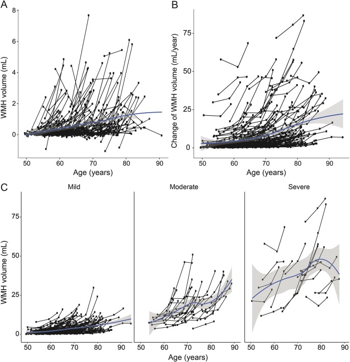
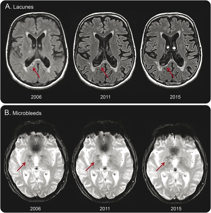
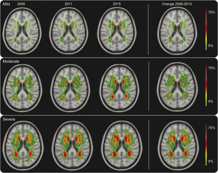
Methods: Changes in SVD markers of 276 participants of the Radboud University Nijmegen Diffusion Tensor and Magnetic Resonance Imaging Cohort (RUN DMC) cohort were assessed at 3 time points over 9 years. We assessed white matter hyperintensities (WMH) volume by semiautomatic segmentation and rated lacunes and microbleeds manually. We categorized baseline WMH severity as mild, moderate, or severe according to the modified Fazekas scale. We performed mixed-effects regression analysis including a quadratic term for increasing age.
Results: Mean WMH progression over 9 years was 4.7 mL (0.54 mL/y; interquartile range 0.95-5.5 mL), 20.3% of patients had incident lacunes (2.3%/y), and 18.9% had incident microbleeds (2.2%/y). WMH volume declined in 9.4% of the participants during the first follow-up interval, but only for 1 participant (0.4%) throughout the whole follow-up. Lacunes disappeared in 3.6% and microbleeds in 5.7% of the participants. WMH progression accelerated over time: including a quadratic term for increasing age during follow-up significantly improved the model (p < 0.001). SVD progression was predominantly seen in participants with moderate to severe WMH at baseline compared to those with mild WMH (odds ratio [OR] 35.5, 95% confidence interval [CI] 15.8-80.0, p < 0.001 for WMH progression; OR 5.7, 95% CI 2.8-11.2, p < 0.001 for incident lacunes; and OR 2.9, 95% CI 1.4-5.9, p = 0.003 for incident microbleeds).
Conclusions: SVD progression is nonlinear, accelerating over time, and a highly dynamic process, with progression interrupted by reduction in some, in a population that on average shows progression.
Copyright © 2017 The Author(s). Published by Wolters Kluwer Health, Inc. on behalf of the American Academy of Neurology.
Figures
References
-
- Prins ND, Scheltens P. White matter hyperintensities, cognitive impairment and dementia: an update. Nat Rev Neurol 2015;11:157–165. - PubMed
-
- Banerjee G, Wilson D, Jager HR, Werring DJ. Novel imaging techniques in cerebral small vessel diseases and vascular cognitive impairment. Biochim Biophys Acta 2016;1862:926–938. - PubMed
-
- Gouw AA, van der Flier WM, van Straaten EC, et al. Reliability and sensitivity of visual scales versus volumetry for evaluating white matter hyperintensity progression. Cerebrovasc Dis 2008;25:247–253. - PubMed
MeSH terms
Grants and funding
LinkOut - more resources
Full Text Sources
Other Literature Sources