. 2017 Oct 27;91(22):e00924-17.
doi: 10.1128/JVI.00924-17. Print 2017 Nov 15.

Foot-and-Mouth Disease (FMD) Virus 3C Protease Mutant L127P: Implications for FMD Vaccine Development

Affiliations

Foot-and-Mouth Disease (FMD) Virus 3C Protease Mutant L127P: Implications for FMD Vaccine Development

Michael Puckette et al. J Virol. .

Abstract

The foot-and-mouth disease virus (FMDV) afflicts livestock in more than 80 countries, limiting food production and global trade. Production of foot-and-mouth disease (FMD) vaccines requires cytosolic expression of the FMDV 3C protease to cleave the P1 polyprotein into mature capsid proteins, but the FMDV 3C protease is toxic to host cells. To identify less-toxic isoforms of the FMDV 3C protease, we screened 3C mutants for increased transgene output in comparison to wild-type 3C using a Gaussia luciferase reporter system. The novel point mutation 3C(L127P) increased yields of recombinant FMDV subunit proteins in mammalian and bacterial cells expressing P1-3C transgenes and retained the ability to process P1 polyproteins from multiple FMDV serotypes. The 3C(L127P) mutant produced crystalline arrays of FMDV-like particles in mammalian and bacterial cells, potentially providing a practical method of rapid, inexpensive FMD vaccine production in bacteria.IMPORTANCE The mutant FMDV 3C protease L127P significantly increased yields of recombinant FMDV subunit antigens and produced virus-like particles in mammalian and bacterial cells. The L127P mutation represents a novel advancement for economical FMD vaccine production.

Keywords: 3C protease; Escherichia coli; Gaussia luciferase; StopGo translation; VLP; adenoviruses; foot-and-mouth disease virus; in vivo; in vivo expression technology; translational interrupter; vaccines; virus-like particles.


Figures

FIG 1
FIG 1
(A) Layout of the bicistronic GLuc-3C reporter assay used to evaluate 3C mutants and protein products. (B) Luciferase readings from culture media of HEK293-T cells transfected with GLuc-3C constructs (data represent an average of 6 samples/3C construct ± standard deviations).
FIG 2
FIG 2
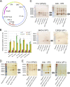
(A) Plasmid construct used for expressing FMDV P1-3C-SGLuc to evaluate P1 processing, transgene expression, and VLP formation. CMV, cytomegalovirus; SV40, simian virus 40. (B) Luciferase readings from culture media of mammalian cells expressing FMDV P1-3C-SGLuc constructs containing the 3C wild type (WT) or C142T, L127P, or C163A mutants (data represent an average of 7 samples/3C construct ± standard deviations [std. dev.]). *, P < 0.001 (for comparisons of luciferase RLU/0.5 s for L127P and C163A by a two-tailed t test). There was significant difference between the RLU/0.5 s values from L127P and those from both the wt and the C142T mutant for each serotype by a two-tailed t test (P < 0.001). (C) Western blots of transfected HEK293-T cell lysates showing P1 products from five FMDV serotypes processed by 3C(L127P). (D) FMDV SAT2 P1-expressing cells analyzed using an anti-VP0/VP2 monoclonal antibody. (E) FMDV A2006 Turkey P1-expressing cells analyzed using anti-VP0/VP2, anti-VP3, and anti-VP1 monoclonal antibodies.
FIG 3
FIG 3
(A to C) TEM images of FMDV VLP arrays in HEK293-T cells transfected with plasmids encoding 3C(L127P) and P1 polyprotein from (A) O1 Manisa, (B) SAT2 Egypt 2012, or (C) Asia1 Shamir. (D and E) FMDV O1 Manisa VLPs emerging from transfected HEK293-T cells (D) and dispersing into individual capsids (E). Bars in panels A and B and on the right side of panels D and E, 100 nm; bar in panel C, 500 nm.
FIG 4
FIG 4
Western blots of lysates from HEK293-T cells transfected with FMDV O1 Manisa P1 and 3C variants (wild-type strain or L127P or C163A mutant). White arrows indicate unprocessed host protein. Gray arrows indicate processed fragments. (A) Western blots showing FMDV P1 polyprotein and processed products of VP0/VP2, VP3, and VP1. (B) Equal levels of loading indicated by anti-GAPDH antibody analysis. (C to G) Blots of 3C protease host protein targets: (C) anti-NEMO; (D) anti-histone H3; (E) anti-eIF4AI; (F) anti-SAM68 N terminus (N-term); (G) anti-SAM68 C terminus (C-term).
FIG 5
FIG 5
Western blot analysis of cell-free reactions with anti-eIF4AI and anti-FLAG antibodies. White arrows indicate unprocessed host protein. Gray arrows indicate processed fragments.
FIG 6
FIG 6
Data from vaccination and challenge study with cattle. (A) Transgene layout of FMDV serotype O PanAsia-2 sequence cloned into Ad5 Blue vector. (B) Number of cattle protected from clinical FMD and viremia by treatment group at 3, 6, 10, and 14 dpc. P = 0.07 (for comparisons of each vaccinated group to the control group; Fisher's exact test). All cattle were challenged 14 dpv with virulent FMDV serotype O PanAsia-2. (C) FMDV serotype O PanAsia-2 virus neutralization test antibody titers (VNT) in serum samples. (D) VNT titers to adenovirus serotype 5 vector. For panels C and D, bars represent the VNT geometric mean titer (GMT) for 4 values (± standard deviations; limit of detection range, >0.6 to 3.6 log10). dpv, days postvaccination; dpc, days postchallenge.
FIG 7
FIG 7
Overnight growth of E. coli bacteria expressing 3C mutants ± the inducer, IPTG.
FIG 8
FIG 8
(A) Diagram of the two-plasmid system used for expression of FMDV P1 and 3C protease in E. coli to produce virus-like particles. (B) Western blot analyses of soluble and insoluble fractions from cell lysates of induced E. coli expressing FMDV O1 Manisa P1 and FLAG-tagged 3C(L127P) demonstrating the presence of processed VP0, VP2, and VP1. (C) TEM image of VLP crystalline arrays in transformed bacteria expressing FMDV O1 Manisa P1 and 3C(L127P). Bar on 20000× panel, 500 nm; bar on 50000× panel, 100 nm.

References

    1. Knight-Jones TJ, Rushton J. 2013. The economic impacts of foot and mouth disease—what are they, how big are they and where do they occur? Prev Vet Med 112:161–173. doi:10.1016/j.prevetmed.2013.07.013. - DOI - PMC - PubMed
    1. Pacheco JM, Brum MC, Moraes MP, Golde WT, Grubman MJ. 2005. Rapid protection of cattle from direct challenge with foot-and-mouth disease virus (FMDV) by a single inoculation with an adenovirus-vectored FMDV subunit vaccine. Virology 337:205–209. doi:10.1016/j.virol.2005.04.014. - DOI - PubMed
    1. Schutta C, Barrera J, Pisano M, Zsak L, Grubman MJ, Mayr GA, Moraes MP, Kamicker BJ, Brake DA, Ettyreddy D, Brough DE, Butman BT, Neilan JG. 2016. Multiple efficacy studies of an adenovirus-vectored foot-and-mouth disease virus serotype A24 subunit vaccine in cattle using homologous challenge. Vaccine 34:3214–3220. doi:10.1016/j.vaccine.2015.12.018. - DOI - PubMed
    1. Mayr GA, Chinsangaram J, Grubman MJ. 1999. Development of replication-defective adenovirus serotype 5 containing the capsid and 3C protease coding regions of foot-and-mouth disease virus as a vaccine candidate. Virology 263:496–506. doi:10.1006/viro.1999.9940. - DOI - PubMed
    1. Doel TR. 2003. FMD vaccines. Virus Res 91:81–99. doi:10.1016/S0168-1702(02)00261-7. - DOI - PubMed

MeSH terms