. 2017 Oct 27;91(22):e01182-17.
doi: 10.1128/JVI.01182-17. Print 2017 Nov 15.

Lysine 242 within Helix 10 of the Pseudorabies Virus Nuclear Egress Complex pUL31 Component Is Critical for Primary Envelopment of Nucleocapsids

Affiliations

Lysine 242 within Helix 10 of the Pseudorabies Virus Nuclear Egress Complex pUL31 Component Is Critical for Primary Envelopment of Nucleocapsids

Sebastian Rönfeldt et al. J Virol. .

Abstract

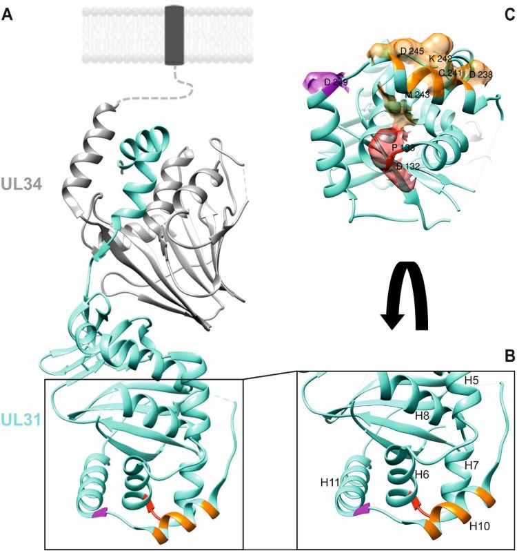
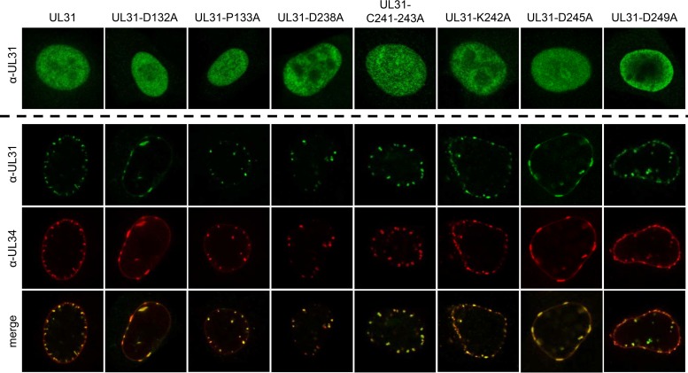
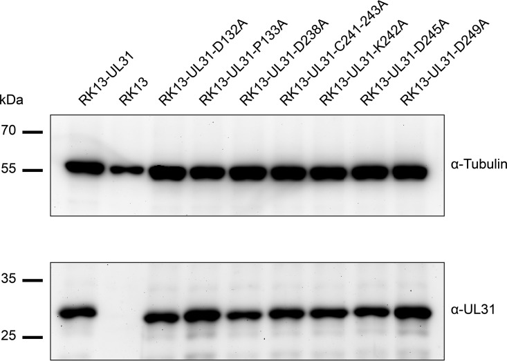
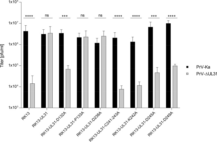
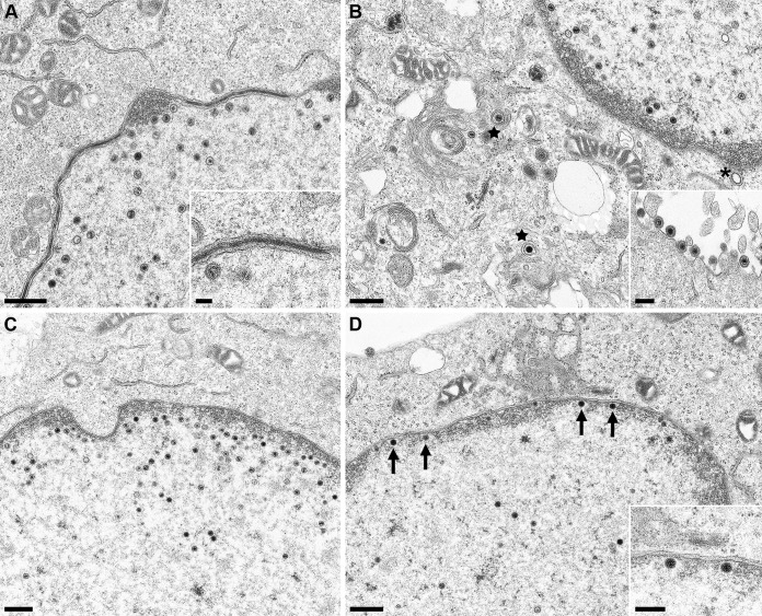
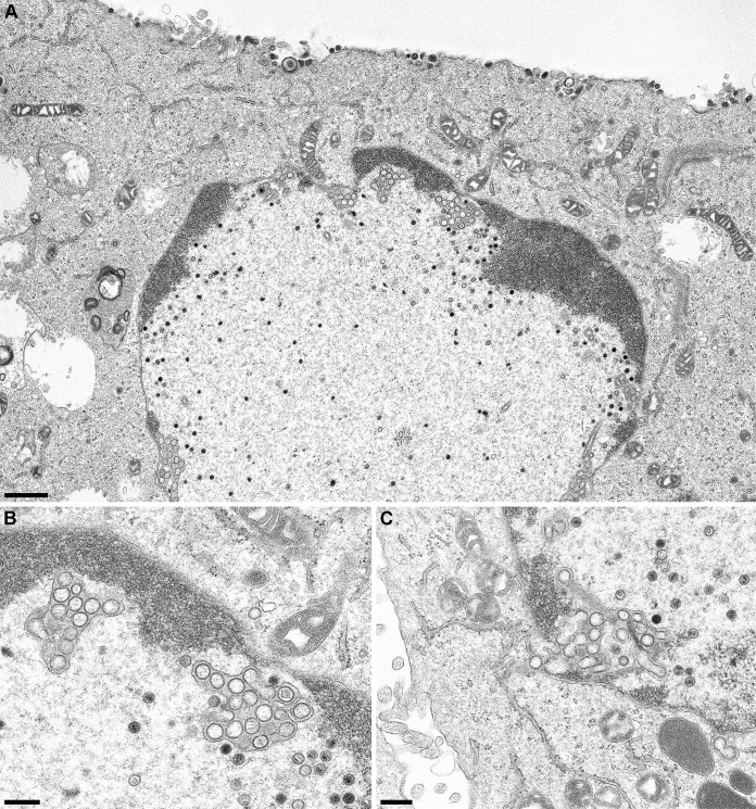
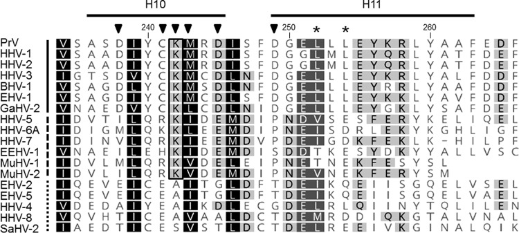
Newly assembled herpesvirus nucleocapsids are translocated from the nucleus to the cytosol by a vesicle-mediated process engaging the nuclear membranes. This transport is governed by the conserved nuclear egress complex (NEC), consisting of the alphaherpesviral pUL34 and pUL31 homologs. The NEC is not only required for efficient nuclear egress but also sufficient for vesicle formation from the inner nuclear membrane (INM), as well as from synthetic lipid bilayers. The recently solved crystal structures for the NECs from different herpesviruses revealed molecular details of this membrane deformation and scission machinery uncovering the interfaces involved in complex and coat formation. However, the interaction domain with the nucleocapsid remained undefined. Since the NEC assembles a curved hexagonal coat on the nucleoplasmic side of the INM consisting of tightly interwoven pUL31/pUL34 heterodimers arranged in hexamers, only the membrane-distal end of the NEC formed by pUL31 residues appears to be accessible for interaction with the nucleocapsid cargo. To identify the amino acids involved in capsid incorporation, we mutated the corresponding regions in the alphaherpesvirus pseudorabies virus (PrV). Site-specifically mutated pUL31 homologs were tested for localization, interaction with pUL34, and complementation of PrV-ΔUL31. We identified a conserved lysine residue at amino acid position 242 in PrV pUL31 located in the alpha-helical domain H10 exposed on the membrane-distal end of the NEC as a key residue for nucleocapsid incorporation into the nascent primary particle.IMPORTANCE Vesicular transport through the nuclear envelope is a focus of research but is still not well understood. Herpesviruses pioneered this mechanism for translocation of the newly assembled nucleocapsid from the nucleus into the cytosol via vesicles derived from the inner nuclear membrane which fuse in a well-tuned process with the outer nuclear membrane to release their content. The structure of the viral nuclear membrane budding and scission machinery has been solved recently, providing in-depth molecular details. However, how cargo is incorporated remained unclear. We identified a conserved lysine residue in the membrane-distal portion of the nuclear egress complex required for capsid uptake into inner nuclear membrane-derived vesicles.

Keywords: NEC capsid interaction; herpesvirus; nuclear egress complex (NEC); nucleocapsid; pUL31; pUL34; pseudorabies virus (PrV).


Figures

FIG 1
FIG 1
Location of putative capsid interaction interfaces in PrV pUL31. (A) In the PrV NEC structure (19), the pUL34 component is shown in gray, and pUL31 is shown in turquoise. Orientation toward and anchorage in the inner nuclear membrane or the primary virion envelope is indicated by the dotted line, and the transmembrane anchor is represented by a dark box. Location of amino acids mutated in this study is indicated in red (D132 and P133), orange (D238, C241, K242, M243, and D245), and magenta (D249). (B) Enlarged membrane-distal end of the NEC in ribbon presentation with alpha-helices numbered, as described previously (19). (C) Amino acids changed in this study are shown in surface presentation using the same coloring. Molecular graphics and analyses were performed with the UCSF Chimera package (35).
FIG 2
FIG 2
Intracellular localization of mutated pUL31 and colocalization with pUL34. Intracellular localization of the mutated proteins was tested after transfection of RK13 cells with the corresponding pUL31 expression plasmids (upper panels). pUL31 was detected with the monospecific anti-pUL31 serum. Speckle formation indicative of vesicle formation from the inner nuclear membrane was analyzed after cotransfection of the indicated pUL31 expression constructs with pcDNA-UL34. pUL31 was detected by the monospecific anti-pUL31 serum (green), and pUL34 was stained with a monoclonal pUL34-specific antibody (red). Merged images are shown in the lowermost panel. Immunofluorescence was recorded with a confocal laser scanning microscope (SP5; Leica, Germany).
FIG 3
FIG 3
pUL31 expression in RK13 cell lines. RK13 cells stably expressing native pUL31 or the mutated proteins, as well as nontransgenic RK13 cells, were harvested by scraping them into the media. Cells were lysed in sample buffer, and proteins were separated in an SDS-10% polyacrylamide gel. After transfer to nitrocellulose, the blot was incubated with the monospecific anti-pUL31 rabbit serum and a monoclonal alpha-tubulin mouse antibody as loading control. Molecular masses of marker proteins are indicated on the left in kilodaltons.
FIG 4
FIG 4
One-step growth. Functional complementation was tested after infection of the cell clones expressing native pUL31 or mutated proteins with PrV-Ka or PrV-ΔUL31 at an MOI of 3. Infected cells and supernatants were harvested at 24 h p.i., and combined titers were determined on RK13-UL31 cells. Nontransgenic RK13 cells were used as a negative control. Shown are mean values of three independent experiments with the corresponding standard deviations. Statistically significant differences are indicated (ns, not significant [P > 0.05]; *, P ≤ 0.05; **, P ≤ 0.01; ***, P ≤ 0.001; ****, P ≤ 0.0001).
FIG 5
FIG 5
Ultrastructural analyses. RK13 cells stably expressing pUL31-D132A (A), pUL31-D238A (B), pUL31-D245A (C), or pUL31-D249A (D) were infected with PrV-ΔUL31 (MOI = 1) and processed for electron microscopy at 14 h p.i. Representative images are shown. Mature capsids in the cytoplasm are marked by stars. A single vesicle in the PNS is indicated by an asterisk (B); nucleocapsids attached to the INM are highlighted by arrows (D). Scale bars: 500 nm in panels A to D, 250 nm in the insets in panels B and D, and 100 nm in the inset in panel A.
FIG 6
FIG 6
Ultrastructural analyses of cells expressing pUL31 mutants defective for capsid incorporation. RK13-UL31-C241-243 cells (A and B) or cells expressing pUL31-K242 (C) were infected as described for Fig. 5. (B) Magnification image of the accumulations of vesicles in the PNS shown in the overview image (panel A). The same accumulations were evident after infection of RK13-UL31-K242 cells (C). Scale bars: 1 μm in panel A and 300 nm in panels B and C.
FIG 7
FIG 7
Amino acid comparison of alpha-helical domains H10 and H11. Amino acid sequences of pUL31 homologs from PrV (AFI70796), HHV-1 (human herpesvirus 1; CAA32324), HHV-2 (CAB06756), HHV-3 (NP_040150), BHV-1 (bovine alphaherpesvirus 1; NP_045327), EHV-1 (equine alphaherpesvirus 1; AAT67286), GaHV-2 (gallid alphaherpesvirus 2; AAF66766), HHV-5 (CAA35412), HHV-6A (NP_042930), HHV-7 (AAC54699), EEHV-1 (elephant endotheliotropic betaherpesvirus; AGE10032), MuHV-1 (murine betaherpesvirus 1; ACE95567), MuHV-2 (AAF99152), EHV-2 (NP_042668), EHV-5 (AIU39595), HHV-4 (AAA45866), HHV-8 (AAB62601), and SaHV-2 (saimiriine gammaherpesvirus 2; Q01041) were aligned using ClustalW alignment (Geneoius version 10.0.9 [36]). Physicochemically similar residues conserved in all sequences aligned are shown by white letters on a black background, those conserved in >80% of the sequences are shown by white letters on a dark gray background, and those conserved in >60% of the sequences are shown in black letters on a gray background. The PrV pUL31 amino acid sequence is shown at the top, with the corresponding numbering and secondary structure elements plotted above the alignment. The lysine residue K242, which is conserved in the alpha- and betaherpesviruses, is boxed. Members of the alphaherpesviruses are marked by a solid line behind the virus abbreviation (left); betaherpesviruses are indicated by a dashed line and gammaherpesviruses by a dotted line. Residues located in H10 and H11 and changed in this study are indicated by a black triangle, and leucine residues mutated in the NES in a previous study (24) are marked by asterisks.

References

    1. Mettenleiter TC. 2002. Herpesvirus assembly and egress. J Virol 76:1537–1547. doi: 10.1128/JVI.76.4.1537-1547.2002. - DOI - PMC - PubMed
    1. Mettenleiter TC, Klupp BG, Granzow H. 2009. Herpesvirus assembly: an update. Virus Res 143:222–234. doi: 10.1016/j.virusres.2009.03.018. - DOI - PubMed
    1. Mettenleiter TC, Muller F, Granzow H, Klupp BG. 2013. The way out: what we know and do not know about herpesvirus nuclear egress. Cell Microbiol 15:170–178. doi: 10.1111/cmi.12044. - DOI - PubMed
    1. Johnson DC, Baines JD. 2011. Herpesviruses remodel host membranes for virus egress. Nat Rev Microbiol 9:382–394. doi: 10.1038/nrmicro2559. - DOI - PubMed
    1. Bigalke JM, Heuser T, Nicastro D, Heldwein EE. 2014. Membrane deformation and scission by the HSV-1 nuclear egress complex. Nat Commun 5:4131. doi: 10.1038/ncomms5131. - DOI - PMC - PubMed

LinkOut - more resources