. 2017 Oct 27;91(22):e00678-17.
doi: 10.1128/JVI.00678-17. Print 2017 Nov 15.

Human Cytomegalovirus Particles Treated with Specific Antibodies Induce Intrinsic and Adaptive but Not Innate Immune Responses

Affiliations

Human Cytomegalovirus Particles Treated with Specific Antibodies Induce Intrinsic and Adaptive but Not Innate Immune Responses

Zeguang Wu et al. J Virol. .

Abstract

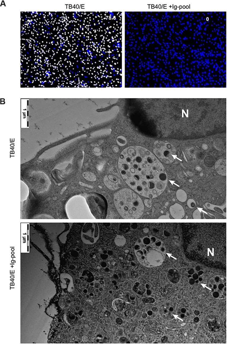
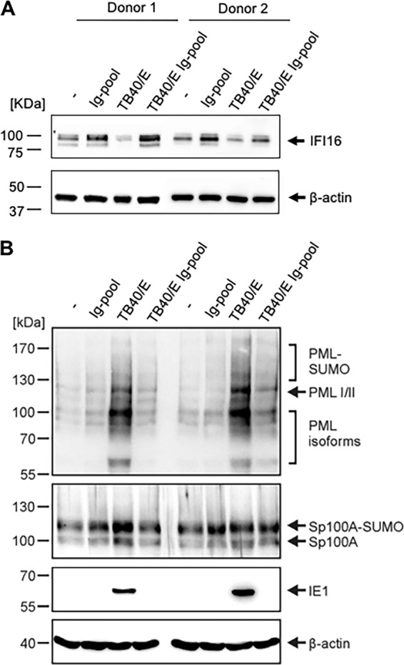
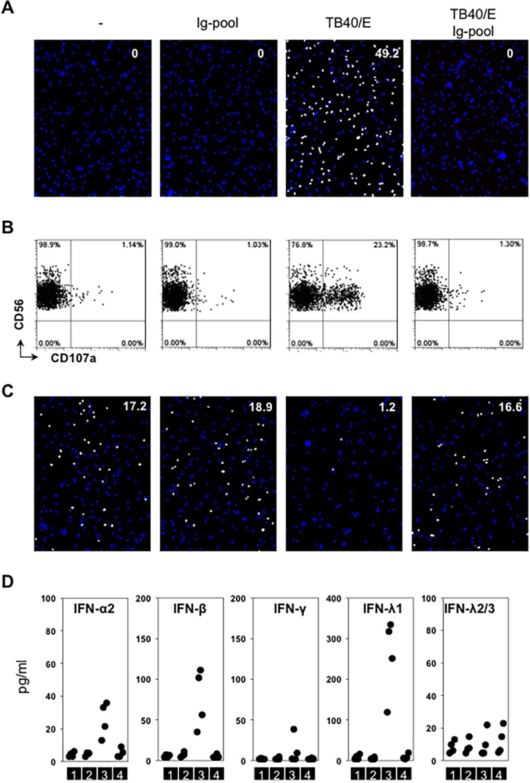
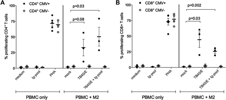
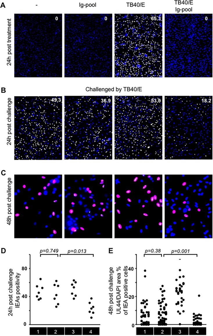
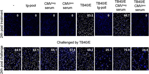
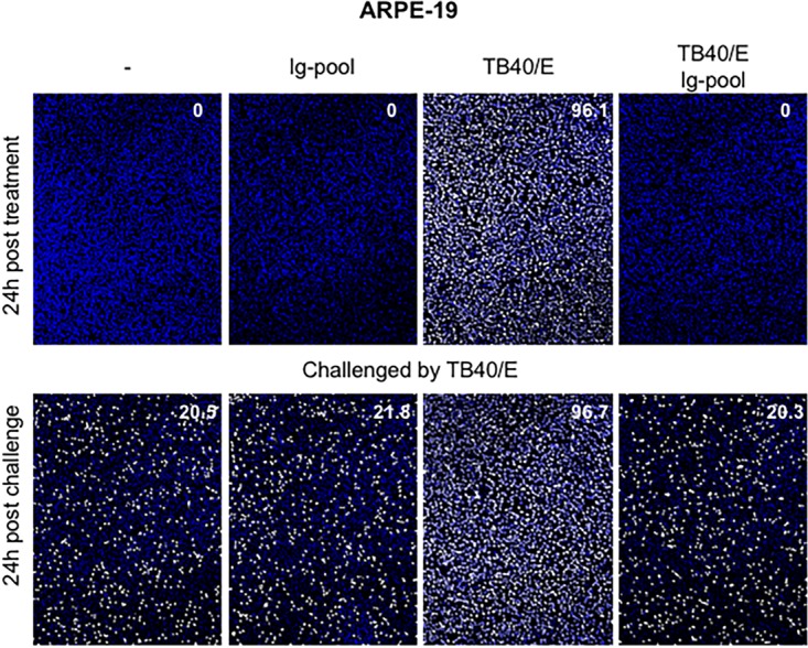
Human cytomegalovirus (HCMV) persistently infects 40% to 100% of the human population worldwide. Experimental and clinical evidence indicates that humoral immunity to HCMV plays an important role in restricting virus dissemination and protecting the infected host from disease. Specific immunoglobulin preparations from pooled plasma of adults selected for high titers of HCMV antibodies have been used for the prevention of CMV disease in transplant recipients and pregnant women. Even though incubation of HCMV particles with these preparations leads to the neutralization of viral infectivity, it is still unclear whether the antibody-treated HCMV particles (referred to here as HCMV-Ab) enter the cells and modulate antiviral immune responses. Here we demonstrate that HCMV-Ab did enter macrophages. HCMV-Ab did not initiate the expression of immediate early antigens (IEAs) in macrophages, but they induced an antiviral state and rendered the cells less susceptible to HCMV infection upon challenge. Resistance to HCMV infection seemed to be due to the activation of intrinsic restriction factors and was independent of interferons. In contrast to actively infected cells, autologous NK cells did not degranulate against HCMV-Ab-treated macrophages, suggesting that these cells may not be eliminated by innate effector cells. Interestingly, HCMV-Ab-treated macrophages stimulated the proliferation of autologous adaptive CD4+ and CD8+ T cells. Our findings not only expand the current knowledge on virus-antibody immunity but may also be relevant for future vaccination strategies.IMPORTANCE Human cytomegalovirus (HCMV), a common herpesvirus, establishes benign but persistent infections in immunocompetent hosts. However, in subjects with an immature or dysfunctional immune system, HCMV is a major cause of morbidity and mortality. Passive immunization has been used in different clinical settings with variable clinical results. Intravenous hyperimmune globulin preparations (IVIg) are obtained from pooled adult human plasma selected for high anti-CMV antibody titers. While HCMV neutralization can be shown in vitro using different systems, data are lacking regarding the cross-influence of IVIg administration on the cellular immune responses. The aim of this study was to evaluate the effects of IVIg on distinct components of the immune response against HCMV, including antigen presentation by macrophages, degranulation of innate natural killer cells, and proliferation of adaptive CD4+ and CD8+ T cells.

Keywords: adaptive immunity; cytomegalovirus; innate immunity; intrinsic defenses; macrophages; neutralizing antibodies.


Figures

FIG 1
FIG 1
HCMV-Ab enter macrophages. A medium containing cell-free TB40/E or a combination of cell-free TB40/E and an Ig pool (TB40/E + IgG-pool) was incubated at 37°C for 30 min. The amount of TB40/E was calculated to reach an MOI of 5. Afterward, the two types of preparations were added to macrophages (1 × 10⁵) in a volume of 100 μl for 1 h. Cells were washed twice and were replenished with new medium. (A) At 24 h posttreatment, macrophages were stained for quantitative evaluation of IEA positive cells (number in the top right corner of the images). The presence of HCMV IEA (white fluorescence) indicates infected cells, and cell nuclei are stained with DAPI (blue fluorescence). Results of one experiment representative of three are shown. (B) At 3 h after treatment, macrophages were washed, fixed by high-pressure freezing, freeze-substituted, plastic embedded, and analyzed by electron microscopy. Round black dots indicate HCMV particles (arrows), and N indicates the nucleus.
FIG 2
FIG 2
IFI16, PML, and Sp100 expression in HCMV-Ab-treated macrophages. Medium alone and medium containing either an Ig pool, cell-free TB40/E, or the combination of cell-free TB40/E and an Ig pool were incubated at 37°C for 30 min. The amount of TB40/E was calculated to reach an MOI of 3. The four media were then added to macrophages (6 × 10⁵) in a volume of 600 μl for 1 h in 24-well plates. Cells were washed twice and were replenished with RMPI 1640 medium. At 24 h posttreatment, the total levels of IFI16 (A), PML, and Sp100 (B) were evaluated by Western blot analysis in cell lysates.
FIG 3
FIG 3
HCMV-Ab do not induce innate immune responses. (A and B) The four types of media described in the legend to Fig. 2 were added to 1 × 10⁵ macrophages seeded in 96-well plates in a volume of 100 μl for 60 min (TB40/E MOI, 3). Cells were washed twice and were incubated in RPMI 1640 medium. After 24 h, treated cells were either stained for quantitative evaluation of IEA positive cells (number in the top right corner of the images) (A) or incubated with autologous purified NK cells (effectors) at an effector-to-target cell ratio of 1 in the presence of an anti-CD107a antibody. NK degranulation was evaluated after 5 h of coculture (B). (C) The supernatants from each condition described for panel A were collected at 24 h posttreatment. They were filtered with a 0.1-μm filter and were transferred to uninfected autologous macrophages (1 × 10⁵) for 24 h. Then the supernatants were discarded, and cells were first challenged with cell-free TB40/E in a volume of 100 μl for 60 min (MOI, 0.3) and then washed twice and incubated in new media. At 24 h postchallenge, IEA expression was evaluated. Results of one experiment representative of four are shown. (D) The supernatants from each condition described for panel A were collected at 24 h posttreatment. The concentrations of interferons in supernatants were tested by bead-based ELISA. Sections 1, 2, 3, and 4 indicate four sequential treatment groups: medium alone, medium containing an Ig pool, medium containing cell-free TB40/E, and medium containing a combination of cell-free TB40/E and an Ig pool.
FIG 4
FIG 4
HCMV-Ab induce adaptive immune responses. Macrophages derived from HCMV-seropositive or -seronegative blood donors (CMV+ and CMV, respectively) were either left untreated (mock), infected with TB40/E at an MOI of 3, or stimulated with TB40/E and an Ig pool. After 1 day, macrophages were harvested, irradiated (30 cGy), and added as stimulators at a ratio of 1:8 to autologous CFSE-labeled PBMC. As controls, PBMC alone were either left untreated (medium) or stimulated with an Ig pool alone or 5 μg/ml phytohemagglutinin (PHA). Six days later, PBMC were harvested and were stained with fluorescently labeled antibodies directed against CD4 and CD8, and the percentage of proliferating CFSElow PBMC was assessed by flow cytometry. Mean values ± standard errors of the means from at least 3 CMV-seronegative and 3 CMV-seropositive blood donors are shown. Each dot represents cells obtained from one blood donor.
FIG 5
FIG 5
HCMV-Ab-treated macrophages are less susceptible to HCMV infection. (A, B, and D) The four types of media described in the legend to Fig. 2 were added to macrophages (1 × 10⁵) in a volume of 100 μl for 60 min (MOI, 3). Cells were washed twice and were incubated in RPMI 1640 medium. After 24 h, treated cells were either stained for quantitative evaluation of IEA positive cells (number in the top right corner of the images) (A) or challenged by cell-free TB40/E in a volume of 100 μl for 60 min at an MOI of 3. Then the cells were washed twice and were replenished with new media. Twenty-four hours later, IEA expression was evaluated (B), and cumulative results from different donors were summarized (D). (C and E) At 48 h postchallenge, the expression of IEA and pUL44 was evaluated. The presence of HCMV IEA (pseudopink fluorescence) indicates infected cells, pUL44 signals are indicated by pseudowhite fluorescence, and cell nuclei are stained blue (DAPI). Ratios of pUL44 fluorescence to nuclear area were analyzed in IEA-positive cells (C), and cumulative results from different donors were summarized (E). Sections 1, 2, 3, and 4 indicate four sequential treatment groups: medium alone, medium containing an Ig pool, medium containing cell-free TB40/E, and medium containing a combination of cell-free TB40/E and an Ig pool.
FIG 6
FIG 6
Serum from CMV-seropositive but not CMV-seronegative subjects is comparable to the IgG pool. Medium alone and media containing either an Ig pool, CMV-seronegative serum, CMV-seropositive serum, cell-free TB40/E, or a combination of cell-free TB40/E with an Ig pool or sera were incubated at 37°C for 30 min prior to inoculation with macrophages. Media were added to macrophages (1 × 10⁵) in a volume of 100 μl for 60 min (MOI, 3). Cells were washed twice and were incubated in RPMI 1640 medium. After 24 h, treated cells were either stained for quantitative evaluation of IEA positive cells (number in the top right corner of the images) or challenged by cell-free TB40/E in a volume of 100 μl for 60 min at an MOI of 3. Then cells were washed twice and were replenished with new medium. Twenty-four hours later, IEA expression was evaluated.
FIG 7
FIG 7
HCMV-Ab-treated epithelial cells are not resistant to HCMV challenge. The four types of media described in the legend to Fig. 2 were added to ARPE-19 retinal pigment epithelial cells (1 × 10⁵) in a volume of 100 μl for 60 min (MOI, 3). Cells were washed twice and were incubated in their respective culture media. After 24 h, treated cells were either stained for quantitative evaluation of IEA positive cells (number in the top right corner of the images) or challenged by cell-free TB40/E in a volume of 100 μl for 60 min at an MOI of 3. Cells were then washed twice and were replenished with new medium. Twenty-four hours later, IEA expression was evaluated by indirect immunofluorescence.

Similar articles

Cited by

References

    1. Pass RF. 2001. Cytomegalovirus, p 2675–2705. In Knipe DM, Howley PM, Griffin DE, Lamb RA, Martin MA, Roizman B, Straus SE (ed), Fields virology, 4th ed, vol 2 Lippincott Williams & Wilkins, Philadelphia, PA.
    1. La Rosa C, Diamond DJ. 2012. The immune response to human CMV. Future Virol 7:279–293. doi:10.2217/fvl.12.8. - DOI - PMC - PubMed
    1. Macagno A, Bernasconi NL, Vanzetta F, Dander E, Sarasini A, Revello MG, Gerna G, Sallusto F, Lanzavecchia A. 2010. Isolation of human monoclonal antibodies that potently neutralize human cytomegalovirus infection by targeting different epitopes on the gH/gL/UL128-131A complex. J Virol 84:1005–1013. doi:10.1128/JVI.01809-09. - DOI - PMC - PubMed
    1. Fouts AE, Chan P, Stephan JP, Vandlen R, Feierbach B. 2012. Antibodies against the gH/gL/UL128/UL130/UL131 complex comprise the majority of the anti-cytomegalovirus (anti-CMV) neutralizing antibody response in CMV hyperimmune globulin. J Virol 86:7444–7447. doi:10.1128/JVI.00467-12. - DOI - PMC - PubMed
    1. Gerna G, Sarasini A, Patrone M, Percivalle E, Fiorina L, Campanini G, Gallina A, Baldanti F, Revello MG. 2008. Human cytomegalovirus serum neutralizing antibodies block virus infection of endothelial/epithelial cells, but not fibroblasts, early during primary infection. J Gen Virol 89:853–865. doi:10.1099/vir.0.83523-0. - DOI - PubMed

Publication types

MeSH terms

Substances