. 2017 Sep 7;2(17):e93344.
doi: 10.1172/jci.insight.93344.

Endothelium-derived extracellular vesicles promote splenic monocyte mobilization in myocardial infarction

Affiliations

Endothelium-derived extracellular vesicles promote splenic monocyte mobilization in myocardial infarction

Naveed Akbar et al. JCI Insight. .

Abstract

Transcriptionally activated monocytes are recruited to the heart after acute myocardial infarction (AMI). After AMI in mice and humans, the number of extracellular vesicles (EVs) increased acutely. In humans, EV number correlated closely with the extent of myocardial injury. We hypothesized that EVs mediate splenic monocyte mobilization and program transcription following AMI. Some plasma EVs bear endothelial cell (EC) integrins, and both proinflammatory stimulation of ECs and AMI significantly increased VCAM-1-positive EV release. Injected EC-EVs localized to the spleen and interacted with, and mobilized, splenic monocytes in otherwise naive, healthy animals. Analysis of human plasma EV-associated miRNA showed 12 markedly enriched miRNAs after AMI; functional enrichment analyses identified 1,869 putative mRNA targets, which regulate relevant cellular functions (e.g., proliferation and cell movement). Furthermore, gene ontology termed positive chemotaxis as the most enriched pathway for the miRNA-mRNA targets. Among the identified EV miRNAs, EC-associated miRNA-126-3p and -5p were highly regulated after AMI. miRNA-126-3p and -5p regulate cell adhesion- and chemotaxis-associated genes, including the negative regulator of cell motility, plexin-B2. EC-EV exposure significantly downregulated plexin-B2 mRNA in monocytes and upregulated motility integrin ITGB2. These findings identify EVs as a possible novel signaling pathway by linking ischemic myocardium with monocyte mobilization and transcriptional activation following AMI.

Keywords: Cardiology; Cell migration/adhesion; Monocytes; Vascular Biology; endothelial cells.


Conflict of interest statement

Conflict of interest: The authors have declared that no conflict of interest exists.

Figures

Figure 1
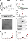
Figure 1. Circulating plasma EV concentration increase with myocardial ischemic injury.
(A) Pearson correlation between the number of plasma extracellular vesicles (EVs, 108/ml) and the degree of ischemic injury in humans (n = 22) in acute myocardial infarction (AMI). (B) Western blot analysis of human plasma EVs for CD9 and CD63. (C) TEM of human plasma EVs. Scale bar: 200 nm. (D and E) Nanoparticle tracking analysis of plasma EVs from wild-type mice subjected to experimental AMI (24 hours) (n = 11–12). (F) Western blot analysis of mouse plasma EVs for ALIX, TSG101, CD63, CD9, Hsp70, ATP5A, histone H3, and CD41. (G) Human (n = 8–24) and (H) mouse (n = 8–13) plasma EV surface markers, CD31, ICAM-1, P selectin, E selectin, and VCAM-1. Values are group mean ± SEM. One-way ANOVA with post-hoc Bonferroni correction or unpaired t test. *P < 0.05, ***P < 0.01.
Figure 2
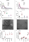
Figure 2. Endothelial cells increase EV release after proinflammatory cytokine stimulation.
(A and B) Nanoparticle tracking analysis (NTA) of human umbilical vein endothelial cell–derived (HUVEC-derived) extracellular vesicles (EVs) released under basal and inflammatory stimulated conditions: TNF-α, IL-1β, IL-4, and IL-6 (n = 5–6). (C) TEM of HUVEC EVs. Scale bar: 200 nm. (D) HUVEC EV surface markers ICAM-1, VCAM-1, P selectin, and E selectin under basal and inflammatory conditions (n = 4–16 per group). (E and F) NTA of sEND.1 EVs released under basal and inflammatory-stimulated conditions: TNF-α, IL-4, and IL-6 (n= 5–11). (G) TEM of sEND.1 EVs. Scale bar: 200 nm. (H) s.END1 EV surface markers ICAM-1, VCAM-1, P selectin, and E selectin under basal and inflammatory conditions (n = 4–8 per group). Values are group mean ± SEM. (A, B, E, and F) One-way ANOVA with post-hoc Bonferroni correction or (D and H) unpaired t test. *P < 0.05, ***P < 0.001.
Figure 3
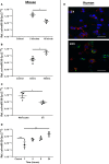
Figure 3. EC-EVs are taken up by monocytes.
(A) Tail vein–injected endothelial cell–derived extracellular vesicles (EC-EVs) transfected with cel-miR39 in mouse blood, (B) spleen, and (C) splenic monocytes (n = 3–5). (D) EC-EVs transfected with cel-miR39 uptake by RAW264.7 cells. (E) PKH67-labeled (green) HUVEC-derived EVs accumulate THP-1 monocyte-derived macrophages (scale bar: 100 μm) (red: F-actin [Phalloidin], nucleus [DAPI: blue]). Group values are 2–Ct (group mean ± SEM). Unpaired 2-tailed t test. *P < 0.05, **P < 0.01.
Figure 4
Figure 4. EC-EVs alter mRNA expression in monocytes.
(A) Plexin-B2 mRNA in THP-1 cells exposed to HUVEC EVs and (B) in RAW264.7 cell exposed to s.END1 EVs. (C) THP-1 cells and (D) RAW264.7 cells show altered relative mRNA expression for ITGB2 after EC-EV exposure. Values are normalized to cyclophilin. Group mean ± SEM 2-ΔCt. Unpaired t test (n = 4–8). *P < 0.05, **P < 0.01.
Figure 5
Figure 5. EC-EVs enhance monocyte chemotaxis.
Inflammatory HUVEC and sEND.1 EVs enhance (A) THP-1 monocyte and (B) RAW264.7 monocyte chemotaxis to MCP-1 (50 nM), respectively, an interaction mediated through VCAM-1 (n = 4–19 per group). Grouped values are mean ± SEM. One-way ANOVA with post-hoc Bonferroni correction. *P < 0.05, **P < 0.01, ***P < 0.001.
Figure 6
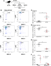
Figure 6. EC-EVs mobilize splenic monocytes.
(A) EC-EVs were tail vein injected into wild-type mice, and monocyte number was quantified by (B) FACS analysis in (C) peripheral blood, (D) bone marrow, and (E) spleen. (F) Monocyte mobilization ratio in mice treated with EC-EVs. Values are the percentage of CD45+ monocytes (mean ± SEM). Unpaired t test (n = 5 per group). **P < 0.01, ***P < 0.001.

References

    1. Ruparelia N, et al. Acute myocardial infarction activates distinct inflammation and proliferation pathways in circulating monocytes, prior to recruitment, and identified through conserved transcriptional responses in mice and humans. Eur Heart J. 2015;36(29):1923–1934. doi: 10.1093/eurheartj/ehv195. - DOI - PMC - PubMed
    1. Swirski FK, et al. Identification of splenic reservoir monocytes and their deployment to inflammatory sites. Science. 2009;325(5940):612–616. doi: 10.1126/science.1175202. - DOI - PMC - PubMed
    1. Panizzi P, et al. Impaired infarct healing in atherosclerotic mice with Ly-6C(hi) monocytosis. J Am Coll Cardiol. 2010;55(15):1629–1638. doi: 10.1016/j.jacc.2009.08.089. - DOI - PMC - PubMed
    1. Nahrendorf M, et al. The healing myocardium sequentially mobilizes two monocyte subsets with divergent and complementary functions. J Exp Med. 2007;204(12):3037–3047. doi: 10.1084/jem.20070885. - DOI - PMC - PubMed
    1. Leuschner F, et al. Therapeutic siRNA silencing in inflammatory monocytes in mice. Nat Biotechnol. 2011;29(11):1005–1010. doi: 10.1038/nbt.1989. - DOI - PMC - PubMed

Publication types

MeSH terms