

SERS-based Immunoassay in a Microfluidic System for the Multiplexed Recognition of Interleukins from Blood Plasma: Towards Picogram Detection
- PMID: 28878312
- PMCID: PMC5587571
- DOI: 10.1038/s41598-017-11152-w
SERS-based Immunoassay in a Microfluidic System for the Multiplexed Recognition of Interleukins from Blood Plasma: Towards Picogram Detection
Abstract
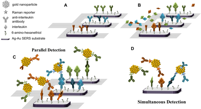
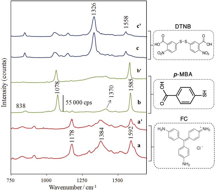
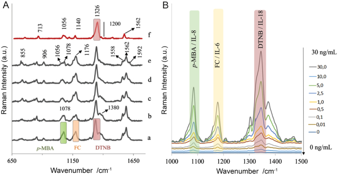
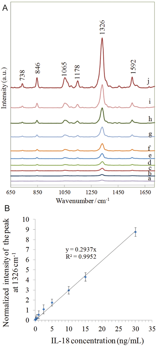
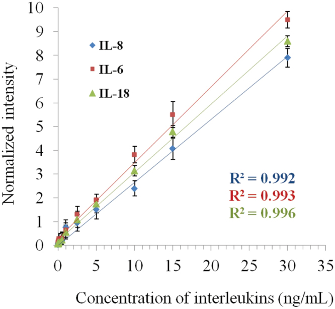
SERS-active nanostructures incorporated into a microfluidic device have been developed for rapid and multiplex monitoring of selected Type 1 cytokine (interleukins: IL-6, IL-8, IL-18) levels in blood plasma. Multiple analyses have been performed by using nanoparticles, each coated with different Raman reporter molecules: 5,5'-dithio-bis(2-nitro-benzoic acid) (DTNB), fuchsin (FC), and p-mercatpobenzoic acid (p-MBA) and with specific antibodies. The multivariate statistical method, principal component analysis (PCA), was applied for segregation of three different antigen-antibody complexes encoded by three Raman reporters (FC, p-MBA, and DTNB) during simultaneous multiplexed detection approach. To the best of our knowledge, we have also presented, for the first time, a possibility for multiplexed quantification of three interleukins: IL-6, IL-8, and IL-18 in blood plasma samples using SERS technique. Our method improves the detection limit in comparison to standard ELISA methods. The low detection limits were estimated to be 2.3 pg·ml-1, 6.5 pg·ml-1, and 4.2 pg·ml-1 in a parallel approach, and 3.8 pg·ml-1, 7.5 pg·ml-1, and 5.2 pg·ml-1 in a simultaneous multiplexed method for IL-6, IL-8, and IL-18, respectively. This demonstrated the sensitivity and reproducibility desirable for analytical examinations.
Conflict of interest statement
The authors declare that they have no competing interests.
Figures
Similar articles
-
Ultrasensitive SERS immunoassay based on diatom biosilica for detection of interleukins in blood plasma.Anal Bioanal Chem. 2017 Nov;409(27):6337-6347. doi: 10.1007/s00216-017-0566-5. Epub 2017 Aug 29. Anal Bioanal Chem. 2017. PMID: 28852782 Free PMC article.
-
Detection of Hepatitis B virus antigen from human blood: SERS immunoassay in a microfluidic system.Biosens Bioelectron. 2015 Apr 15;66:461-7. doi: 10.1016/j.bios.2014.10.082. Epub 2014 Nov 13. Biosens Bioelectron. 2015. PMID: 25497986
-
Simultaneous and highly sensitive detection of multiple breast cancer biomarkers in real samples using a SERS microfluidic chip.Talanta. 2018 Oct 1;188:507-515. doi: 10.1016/j.talanta.2018.06.013. Epub 2018 Jun 6. Talanta. 2018. PMID: 30029406
-
A new protein A assay based on Raman reporter labeled immunogold nanoparticles.Biosens Bioelectron. 2008 Oct 15;24(2):178-83. doi: 10.1016/j.bios.2008.03.035. Epub 2008 Apr 4. Biosens Bioelectron. 2008. PMID: 18468881
-
Analytical characterization using surface-enhanced Raman scattering (SERS) and microfluidic sampling.Nanotechnology. 2015 Mar 6;26(9):092001. doi: 10.1088/0957-4484/26/9/092001. Nanotechnology. 2015. PMID: 25676092 Review.
Cited by
-
Recent Progresses in Optical Biosensors for Interleukin 6 Detection.Biosensors (Basel). 2023 Sep 21;13(9):898. doi: 10.3390/bios13090898. Biosensors (Basel). 2023. PMID: 37754132 Free PMC article. Review.
-
Modulating the interaction of graphenic substrates with human interleukin-6 and its monoclonal antibody: a study by Raman images.RSC Adv. 2023 May 17;13(22):15114-15120. doi: 10.1039/d3ra01627g. eCollection 2023 May 15. RSC Adv. 2023. PMID: 37207094 Free PMC article.
-
Emerging Technologies and Platforms for the Immunodetection of Multiple Biochemical Markers in Osteoarthritis Research and Therapy.Front Med (Lausanne). 2020 Oct 21;7:572977. doi: 10.3389/fmed.2020.572977. eCollection 2020. Front Med (Lausanne). 2020. PMID: 33195320 Free PMC article. Review.
-
Multiplexed Immunosensors and Immunoarrays.Anal Chem. 2020 Jan 7;92(1):345-362. doi: 10.1021/acs.analchem.9b05080. Epub 2019 Dec 2. Anal Chem. 2020. PMID: 31726821 Free PMC article. Review. No abstract available.
-
Recent Advances in Sandwich SERS Immunosensors for Cancer Detection.Int J Mol Sci. 2022 Apr 25;23(9):4740. doi: 10.3390/ijms23094740. Int J Mol Sci. 2022. PMID: 35563131 Free PMC article. Review.
References
Publication types
MeSH terms
Substances
LinkOut - more resources
Full Text Sources
Other Literature Sources
Miscellaneous
