. 2017 Sep 6;8(1):464.
doi: 10.1038/s41467-017-00489-5.

Enoyl-CoA hydratase-1 regulates mTOR signaling and apoptosis by sensing nutrients

Affiliations

Enoyl-CoA hydratase-1 regulates mTOR signaling and apoptosis by sensing nutrients

Ya-Kun Zhang et al. Nat Commun. .

Abstract

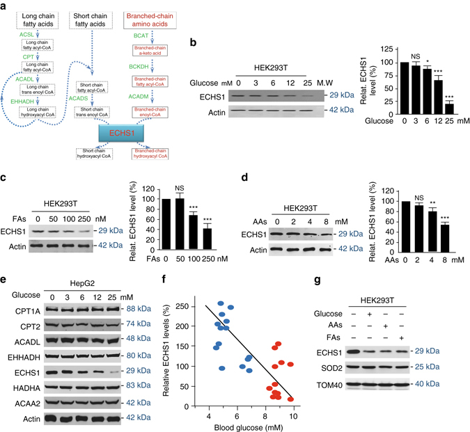
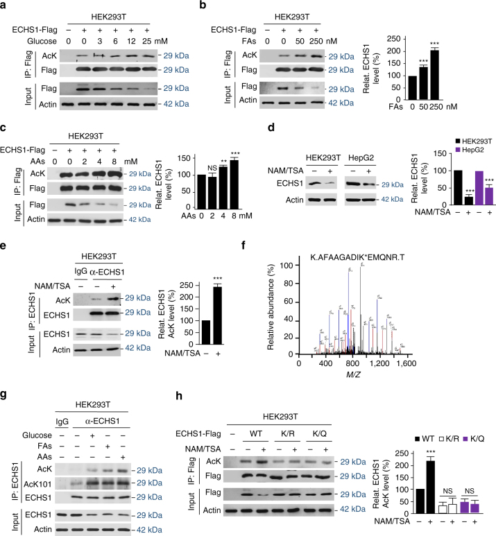
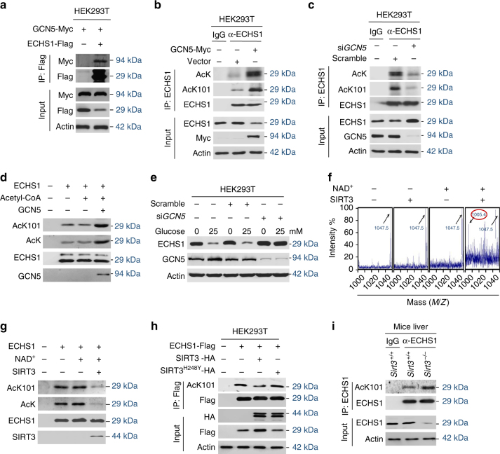
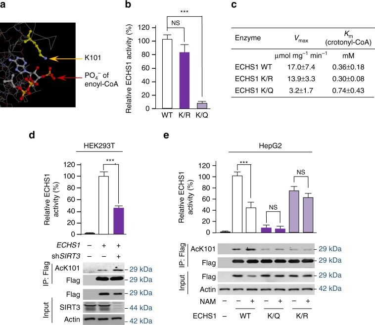
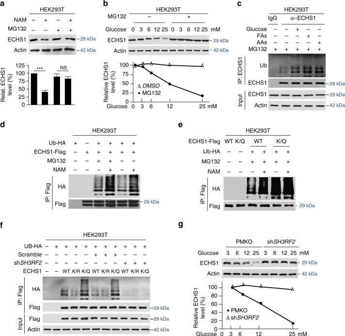
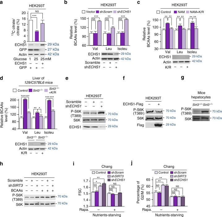
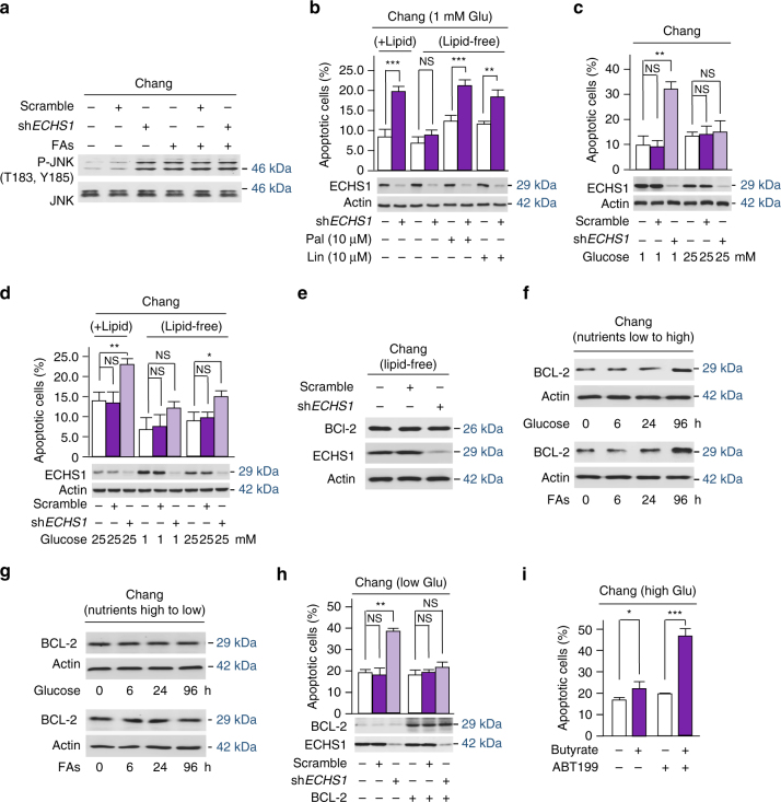
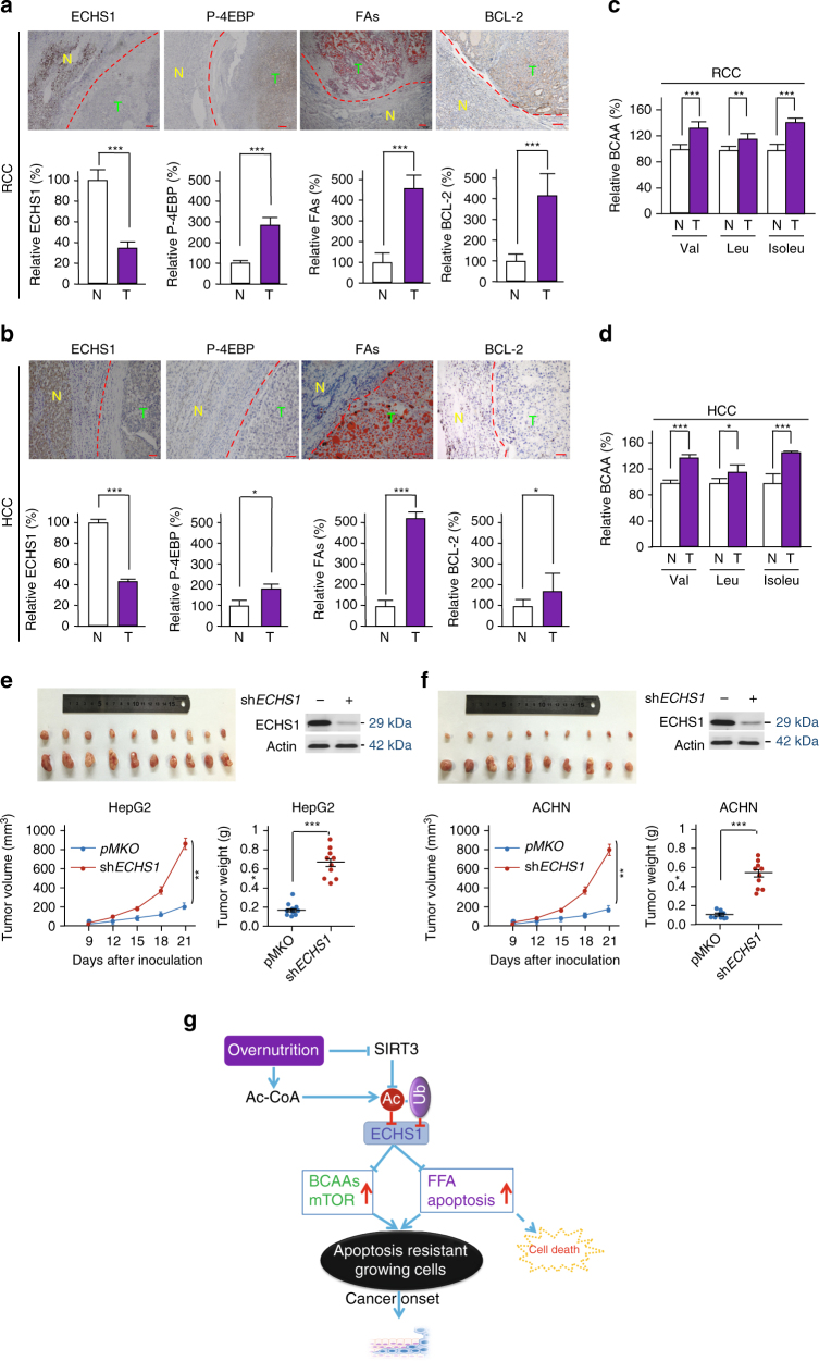
The oncogenic mechanisms of overnutrition, a confirmed independent cancer risk factor, remain poorly understood. Herein, we report that enoyl-CoA hydratase-1 (ECHS1), the enzyme involved in the oxidation of fatty acids (FAs) and branched-chain amino acids (BCAAs), senses nutrients and promotes mTOR activation and apoptotic resistance. Nutrients-promoted acetylation of lys101 of ECHS1 impedes ECHS1 activity by impairing enoyl-CoA binding, promoting ECHS1 degradation and blocking its mitochondrial translocation through inducing ubiquitination. As a result, nutrients induce the accumulation of BCAAs and FAs that activate mTOR signaling and stimulate apoptosis, respectively. The latter was overcome by selection of BCL-2 overexpressing cells under overnutrition conditions. The oncogenic effects of nutrients were reversed by SIRT3, which deacetylates lys101 acetylation. Severely decreased ECHS1, accumulation of BCAAs and FAs, activation of mTOR and overexpression of BCL-2 were observed in cancer tissues from metabolic organs. Our results identified ECHS1, a nutrients-sensing protein that transforms nutrient signals into oncogenic signals.Overnutrition has been linked to increased risk of cancer. Here, the authors show that exceeding nutrients suppress Enoyl-CoA hydratase-1 (ECHS1) activity by inducing its acetylation resulting in accumulation of fatty acids and branched-chain amino acids and oncogenic mTOR activation.


Conflict of interest statement

The authors declare no competing financial interests.

Figures

Fig. 1
Fig. 1
ECHS1 protein levels are decreased by nutrients. a Schematic diagram of catabolic pathways of FA (black-colored) and BCAA (red-colored) oxidation that involve ECHS1. Long chain FAs are oxidized to short-chained intermediates prior to being completely oxidized through ECHS1. BCAAs are oxidized through ECHS1. ACADL acyl-CoA dehydrogenase, long chain, ACADM medium-chain specific acyl-CoA dehydrogenase, mitochondrial, ACADS short-chain specific acyl-CoA dehydrogenase, ACSL acyl-CoA synthetase long-chain, BCAT branched-chain amino-acid transaminase, BCKDH branched-chain keto acid dehydrogenase, carnitine palmitoyltransferase, ECHS1 enoyl CoA hydratase, short chain 1, mitochondrial, EHHADH Enoyl-CoA, Hydratase/3-Hydroxyacyl CoA Dehydrogenase. bd Endogenous ECHS1 levels were detected in HEK293T cells treated with different concentrations of glucose (b), FAs (linoleic acid + palmitic acid) (c), and amino acids (glutamate + aspartate) (d). Representative western blot results and quantitation (herein after) of triplicated western blot are shown. e HepG2 cells were exposed to indicated concentrations of glucose. Levels of endogenous β-oxidation enzymes were detected 4 h after glucose exposure. CPT1A Carnitine O-palmitoyltransferase 1, liver isoform, Carnitine O-palmitoyltransferase 2, mitochondrial, HADHA Trifunctional enzyme subunit alpha, mitochondrial, ACAA2 3-ketoacyl-CoA thiolase, mitochondrial. f Plasma ECHS1 levels of normal people (blue, n = 14) and untreated patients with diabetes (red, n = 12) were determined by western blots. Relative ECHS1 levels (normalized to the average ECHS1 level) were plotted against serum glucose levels. g Mitochondria of HEK293T cells cultured in DMEM base, DMEM base supplemented with glucose (25 mM), fatty acids (250 nM) and amino acids (8 mM), respectively, were isolated and the relative (to SOD2 and TOM40) mitochondrial ECHS1 levels were compared. For bd, mean values of quantitation with SD are reported. *P < 0.05; **P < 0.01; ***P < 0.001
Fig. 2
Fig. 2
Nutrients promote ECHS1 K101 acetylation. ac Western blot detection of acetylation levels of ectopically expressed ECHS1 in HEK293T cell cultured in the presence of excessive glucose (a), FAs (b), and amino acids (c) in the culture media. d Levels of endogenous ECHS1 in HEK293T and HepG2 cells were compared with and without the presence of deacetylases inhibitors NAM/TSA. e Effects of deacetylase inhibitors on the acetylation levels of affinity-purified endogenous ECHS1. f The MS spectrum that led to the identification of K101 acetylation containing tryptic peptide from endogenous ECHS1. g Western blot detection of proteins levels, AcK levels and AcK101 levels of endogenous ECHS1 in HEK293T cells that were cultured in DMEM base and DMEM base supplemented with glucose (25 mM), fatty acids (250 nM), and amino acids (8 mM). h Flag-tagged wildtype, K101R (K/R), and K101Q (K/Q) ECHS1 were overexpressed in HEK293T cells that were cultured in media with or without NAM/TSA. The relative acetylation levels of ECHS1 proteins purified from the above mentioned cells using Flag beads was determined. For be and h, mean values of quantitation with SD are reported. *P < 0.05; **P < 0.001; ***P < 0.001
Fig. 3
Fig. 3
GCN5 acetylates and SIRT3 deacetylates ECHS1. a Myc-tagged GCN5 and Flag-tagged ECHS1 were co-expressed in HEK293T cells. The co-purified Myc-tagged GCN5 was detected in Flag beads purified proteins. b, c Western blot detection of endogenous ECHS1, and AcK and AcK101 levels of affinity-purified ECHS1 from HEK293T cells and GCN5 overexpressing HEK293T cells (b) and from HEK293T cells and GCN5 knockdown HEK293T cells (c). d Western blot detection of AcK and AcK101 levels of recombinant ECHS1 that before and after in vitro acetylation by GCN5. e The levels of endogenous ECHS1 were detected in HEK293T and GCN5 knockdown HEK293T cells that were cultured in the absence of glucose or presence of glucose. f The synthetic acetylated K101-containing ECHS1 peptide was deacetylated by recombinant SIRT3. The synthetic peptide (1047.5) and de-acetylated products (1005.4, red-circled) were assayed by MS. g The levels of AcK and AcK101 of hyperacetylated ECHS1 (from NAM treated cells) were detected before and after in vitro treatment by SIRT3. h ECHS1 was co-expressed with SIRT3 or SIRT3H248Y in HEK293T cells. The AcK101 levels of purified ECHS1 were determined. i The Ac101 levels of affinity-purified ECHS1 from liver of Sirt3 KO and control mice
Fig. 4
Fig. 4
Acetylation inactivates ECHS1. a The crystal structure shows that K101 is involved in enoyl-CoA binding. Arrows point to the amine on lysine (yellow) and the phosphate on enoyl-CoA (red). b The relative specific activities of ECHS1 wild-type (set as 100%), K/R, and K/Q mutants were determined. Mean values with SD are reported. NS not significant; ***P < 0.001. c The V max and K m were determined for wild type ECHS1, K/R, and K/Q mutant using crotonyl-CoA as the substrate. d The activities of ECHS1 expressed in HEK293T or SIRT3 knocked down HEK293T cells were determined. e The K101 acetylation levels and relative specific activities of Flag-tagged wild type, K/Q, and K/R mutant ECHS1 expressed in HepG2 cells cultured in the presence or absence of NAM (5 mM). The average specific activities (n = 3) with SD and representative western blots are shown
Fig. 5
Fig. 5
K101 acetylation promotes ECHS1 ubiquitination and degradation. a NAM treatment effects on endogenous ECHS1 levels in HEK293T cells were analyzed in the presence and absence of MG132. Mean values of quantitation with SD are reported. NS not significant; ***P < 0.001. b The endogenous ECHS1 levels in HEK293T cells that were cultured under various glucose levels were detected under absence (DMSO) and presence treatments with MG132. c The ubiquitination of affinity-purified endogenous ECHS1 from HEK293T cells that were cultured with DMEM base and DMEM base supplemented with glucose, fatty acids, and amino acids, respectively, were determined. d Flag-tagged ECHS1 was expressed in HEK293T cells. The ubiquitination levels of ECHS1 expressed from cells cultured with either NAM or MG132, or both NAM and MG132 treatments were analyzed. e Flag-tagged ECHS1 or K/Q mutant were expressed alone or with HA-tagged ubiquitin in HEK293T cells cultured with or without NAM. The ubiquitination levels of Flag bead affinity-purified ECHS1 were analyzed. f The ubiquitination levels of ectopically expressed Flag-tagged ECHS1, K/R and K/Q mutants from HEK293T and shSH3RF2 knockdown (KD effects please see Supplementary Fig. 7d) HEK293T cells. g Endogenous ECHS1 levels were detected in HEK293T cells and SH3RF2 knocked down (KD effects please see Supplementary Fig. 7d) HEK293T cells that were cultured in media contained indicated glucose levels. Relative ECHS1 levels were all normalized to that of glucose of 3 mM
Fig. 6
Fig. 6
K101 acetylation inhibits ECHS1 mitochondria translocation. a The K101 acetylation levels of ECHS1 in the cytosol, mitochondria membrane, and mitochondria matrix were detected after mouse primary hepatocytes were exposed to 25 mM glucose for 0, 2, 4, 6, and 8 h. b Flag-tagged ECHS1 was co-expressed with HA-tagged ubiquitin in HEK293T cells. ECHS1 ubiquitination levels in the cytosol, mitochondria membrane, and mitochondria matrix were detected under low and high glucose treatment for 4 h. GAPDH, COX IV and SOD2 were used as markers of cytosol, mitochondria membrane and mitochondria matrix, respectively. c ECHS1 levels in the cytosol, mitochondria membrane, and mitochondria matrix were analyzed (n = 3) after cells were exposed to 25 mM glucose for 3, 6, 9, and 12 h. Representative western blots are presented. All levels were normalized to those of cells at time 0 and the average relative levels were quantified (bottom). d Schematic diagram of ECHS1 regulation by nutrients. Nutrient-induced acetylation inactivates ECHS1 and induces ubiquitination of ECHS1 that blocks ECHS1 mitochondrial translocation and promotes ECHS1 degradation. These effects synergistically inactivate ECHS1
Fig. 7
Fig. 7
Nutrient activates mTOR signaling by accumulating BCAAs. a The conversion of Leucine-1,2-13C2 to 13C-labeled citrate was traced by targeted liquid-chromatography tandem mass spectrometry (LC–MS/MS) in HEK293T cells cultured in low (1 mM) and high (25 mM) glucose media, and in ECHS1 overexpressing HEK293T cells cultured in high glucose media. b The BCAA levels in HEK293T cells and ECHS1 knocked down HEK293T cells were determined (n = 4). BCAA levels were normalized to those of HEK293T cells. c BCAA levels of HEK293T cells, NAM treated HEK293T cells, and NAM treated HEK293T cells with overexpression of ECHS1K101R were determined by GC–MS (n = 4). All levels were normalized to those of HEK293T cells. The expression of ECHS1K/R was confirmed by western blot. d BCAAs levels in the liver of 129/C57BL6 mice, Sirt3 knockout 129/C57BL6 mice, and ECHS1K101R overexpressing Sirt3 knockout 129/C57BL6 mice were determined (n = 6). ECHS1K/R expression was achieved by tail vein injection of the ECHS1K/R plasmid, and the expression was confirmed by western blot. BCAA levels were normalized to those in the liver of isogenic control mice. e The levels of P-T389 on S6K were determined in HEK293T cells with and without ECHS1 knockdown. f The levels of P-T389 on S6K were determined in HEK293T cells with and without ECHS1 overexpression. g The levels of P-T389 on S6K were determined in the hepatocytes of Sirt3 KO and its isogenic control mice. h The effects of shSIRT3 knockdown (for knockdown effects see Figure 4) on P-T389 of S6K were detected in HEK293T cells cultured with or without BCAA supplementation in the medium. i The cell size of Chang cells and ECHS1 or SIRT3 knocked down Chang cells were determined by measuring forward side scatter (FSC) in the presence or absence of rapamycin. j The percentage of Chang cells and ECHS1 or SIRT3 knocked down Chang cells in the G2/M phase was determined by fluorescence-activated cell sorter (FACS) in the presence or absence of rapamycin. For all figures, mean values with SD are reported. NS not significant; *P < 0.05; **P < 0.01; ***P < 0.001
Fig. 8
Fig. 8
Nutrient induces FAs accumulation. a The conversion of 13C-palmitate to 13C-citrate was traced in HEK293T cells cultured in low and high glucose media, and in ECHS1 overexpressing HEK293T cells cultured in high glucose media. b Relative FAs levels in HEK293T cells, ECHS1 knocked down HEK293T cells and ECHS1 knocked down HEK293T cells overexpressing shRNA resistant ECHS1 (ECHS1-r) were detected. All levels were normalized to the total proteins. c The relative butyrate levels in Chang liver cells and ECHS1 knocked down Chang liver cells were determined (n = 4). The butyrate levels in untreated Chang liver cells (25 μM) were set as 100%. d The relative FAs levels in HEK293T cells cultured in low glucose (−), high glucose (+), and high glucose with ECHS1K101R overexpression were determined (n = 3). e The FAs levels in the liver of wild type, Sirt3 −/−, and ECHS1 K/R overexpressing Sirt3 −/− 129/C57BL6 mice were assessed by oil red staining. Four mice of each group were analyzed. Relative FAs levels were determined by comparing intensity of signals, Sirt3 +/+ value was set as 100% arbitrarily (right). f The relative (to time 0) intracellular FAs levels in Chang cells that were maintained in high glucose (25 mM) and low glucose (1 mM) were detected (n = 3) at time points as indicated. For all figures, mean values with SD are reported. NS not significant; *P < 0.05; **P < 0.01; ***P < 0.001
Fig. 9
Fig. 9
Nutrient induces apoptosis resistance. a The p-T183 and p-Y185 levels of JNK were analyzed for their responses to ECHS1 knockdown in Chang cells cultured in DMEM media with and without fatty acids supplementation. b The apoptotic rates of Chang and ECHS1 knockdown Chang cells that were cultured under presence and absence of lipids, and lipid-free media supplemented with saturated palmitate or unsaturated linoleic acid, were determined (n = 4). c The percentages of apoptotic Chang cells and apoptotic ECHS1 knocked down Chang cells were determined when cells were cultured in the presence of 1 mM and 25 mM glucose (n = 4). d The apoptotic rates of Chang and ECHS1 knockdown Chang cells that were cultured under presence and absence of lipids, and lipid-free media supplemented with low and high glucose, were determined (n = 4). e BCL-2 levels in Chang cells and ECHS1 knockdown Chang cells that were cultured in lipid-free media were compared. f Chang cells were conditioned in glucose- or fatty acids-free media for 24 h before they were transferred to media containing high glucose (25 mM) or high fatty acids (250 nM), BCL2 levels were monitored over time. g Chang cells were conditioned in high glucose or high fatty acids for 96 h before they were transferred to low glucose (1 mM) or low fatty acids (250 nM) media, BCL2 levels were monitored over time. h The apoptotic rates in Chang cells and ECHS1 knockdown Chang cells cultured under low glucose were determined under with and without BCL-2 overexpression. i Chang cells were cultured in the presence of 25 mM glucose. The percentages of apoptotic cells of untreated and 1 mM butyrate-treated cells with or without ABT-199 treatment were determined (n = 4). For all figures, mean values with SD are reported. not significant; *P < 0.05; **P < 0.001; ***P < 0.001
Fig. 10
Fig. 10
ECHS1 downregulation promotes BCAA and FA accumulation and cancer growth. a, b IHC analysis for ECHS1, P-4EBP, FAs, and BCL-2 in RCC (a, scale bars: 200 μm) and HCC (b, scale bars: 50 μm) specimens and their respective adjacent normal tissues. c, d Relative levels of BCAAs were determined in RCC (c) and HCC (d) tissues and their respective adjacent normal tissues. The levels of BCAAs in normal tissues were set as 100%. e, f The xenograft tumor growth of HepG2, ECHS1 knockdown HepG2, ACHN and ECHS1 knockdown ACHN cells were analyzed in Balb/c nude mice (4–6 weeks old). g Schematic diagram indicating how rich nutrients enrich ECHS1-inactivated cells and promote cancer initiation. Nutrients inactivate ECHS1 cause accumulation of BCAAs and FFAs, which induce mTOR activation and lipoapoptosis, respectively. The latter resulted in selection for apoptosis resistant cells and facilitate cancer transformation. Mean values with SD are reported. *P < 0.05; **P < 0.01; ***P < 0.001

References

    1. Moore JB. Symposium 1: overnutrition: consequences and solutions non-alcoholic fatty liver disease: the hepatic consequence of obesity and the metabolic syndrome. Proc. Nutr. Soc. 2010;69:211–220. doi: 10.1017/S0029665110000030. - DOI - PubMed
    1. Yam D. Insulin-cancer relationships - possible dietary implication. Med. Hypotheses. 1992;38:111–117. doi: 10.1016/0306-9877(92)90082-N. - DOI - PubMed
    1. Benjamin D, Colombi M, Moroni C, Hall MN. Rapamycin passes the torch: a new generation of mTOR inhibitors. Nat. Rev. Drug Discov. 2011;10:868–880. doi: 10.1038/nrd3531. - DOI - PubMed
    1. Zoncu R, Efeyan A, Sabatini DM. mTOR: from growth signal integration to cancer, diabetes and ageing. Nat. Rev. Mol. Cell Biol. 2011;12:21–35. doi: 10.1038/nrm3025. - DOI - PMC - PubMed
    1. Yeung SJ, Pan J, Lee MH. Roles of p53, MYC and HIF-1 in regulating glycolysis - the seventh hallmark of cancer. Cell. Mol. Life Sci. 2008;65:3981–3999. doi: 10.1007/s00018-008-8224-x. - DOI - PMC - PubMed

Publication types

MeSH terms