Review
. 2017 Oct;18(10):573-584.
doi: 10.1038/nrn.2017.107. Epub 2017 Sep 7.

The use of brain organoids to investigate neural development and disease

Affiliations
Review

The use of brain organoids to investigate neural development and disease

Elizabeth Di Lullo et al. Nat Rev Neurosci. 2017 Oct.

Abstract

Understanding the development and dysfunction of the human brain is a major goal of neurobiology. Much of our current understanding of human brain development has been derived from the examination of post-mortem and pathological specimens, bolstered by observations of developing non-human primates and experimental studies focused largely on mouse models. However, these tissue specimens and model systems cannot fully capture the unique and dynamic features of human brain development. Recent advances in stem cell technologies that enable the generation of human brain organoids from pluripotent stem cells (PSCs) promise to profoundly change our understanding of the development of the human brain and enable a detailed study of the pathogenesis of inherited and acquired brain diseases.


Conflict of interest statement

Competing interests statement

The authors declare no competing interests.

Figures

Figure 1
Figure 1. Cortical organoids generated with current protocols
Schematic representation of cortical organoids generated with current protocols. Immunohistochemical analyses reveal rosette-like structures in immature organoids (left). These contain neuroepithelial stem cells and ventricular radial glial cells (vRGs) that divide at the apical surface and form a ventricular-like zone (VZ). Intermediate progenitors (IPs) and neurons surround the VZ. Cells that express markers of early cortical plate neurons such as COUP-TF-interacting protein 2 (CTIP2, also known as BCL11B) and T-box brain protein 1 (TBR1) are also generated in immature organoids,,. More mature organoids (right) display multiple progenitor zones, including a VZ and a subventricular-like zone (SVZ). Immunohistochemistry reveals the presence of outer radial glial cells (oRGs), forming the outer subventricular zone (OSVZ) and the presence of cells expressing specific cortical layer markers and glial cell markers. The molecular markers of cell identity demonstrated in this schematic are based on findings from REF . BRN2, POU domain, class 3, transcription factor 2; CUX1, homeobox protein cut-like 1; GFAP, glial fibrillary acidic protein; HOPX, homeodomain-only protein; PAX6, paired box protein Pax-6.
Figure 2
Figure 2. Human cerebral organoids as models of cortical development
a | Schematics illustrating the cellular composition and laminar organization of the developing human and mouse cortex and that of a typical human organoid (not to scale),,. Human cortex and human organoid models notably share an expanded subventricular zone (SVZ) that contains an outer SVZ (OSVZ),. Human organoid models therefore provide an important advance in our ability to study the role of the expanded human SVZ and the cell types associated with this region in vitro. Outer radial glial cells (oRGs), characteristic of the human SVZ, have been identified in organoids,,,,. Over time, cortical organoids are able to generate diverse neuronal subtypes that can become organized into deep and upper layers as has been shown by immunohistochemistry,,,. However, current models do not display the complexity or organization of mouse or human laminar organization. b | Current organoid protocols do not produce all the cell types known to be important for human cortical development. The schematic illustrates some of the cell types under-represented in current organoid models. Importantly, cerebral organoids lack vascularization (endothelial cells) and microglial cells. Oligodendrocyte precursor cells (OPCs) are also uncommon in most protocols. CP, cortical plate; IP, intermediate progenitor cell; IZ, intermediate zone; MZ, marginal zone; SP, subplate; vRG, ventricular radial glial cell.
Figure 3
Figure 3. Aspects of human cortical development for future exploration in brain organoid models
Several interesting aspects of human cortical development are yet to be explored in brain organoid models. a | A developmental switch from a continuous radial glial scaffold to a discontinuous scaffold and the generation of radial glial cells with non-classical morphologies (such as truncated radial glial cells (tRGs)) was recently described and could be examined in organoid models. It was shown that during early neurogenesis (the continuous scaffold stage), the basal fibres of ventricular radial glial cells (vRGs) contact the pial surface and that newborn neurons migrate along the fibres of both vRGs and outer radial glial cells (oRGs). During late neurogenesis (the discontinuous scaffold stage), newborn neurons reach the cortical plate only along oRG fibres. If these structures are recapitulated in human organoids, time-lapse imaging of migrating neurons could be used to demonstrate this developmental switch. b | Another feature of human cortical development to be explored is the cortical folding that takes place largely after neurogenesis is complete, which has yet to be properly modelled in organoids. In addition, the establishment of correct lamination replicating the six layers (I–VI) of the mammalian cortex has not yet been replicated in organoids. Extracortical input and canonical intracortical circuits, including those mediating inhibition, have not yet been fully demonstrated in organoids, and the roles of extrinsic signalling via morphogens and other diffusible cues remain largely unexplored. IP, intermediate progenitor cell. Part a adapted with permission from REF. , Elsevier.

References

    1. Tiscornia G, Vivas EL, Belmonte JCI. Diseases in a dish: modeling human genetic disorders using induced pluripotent cells. Nat Med. 2011;17:1570–1576. - PubMed
    1. Lancaster MA, et al. Cerebral organoids model human brain development and microcephaly. Nature. 2013;501:373–379. This was the first paper to introduce a method for generating cerebral organoids. The organoids were used to model microcephaly. - PMC - PubMed
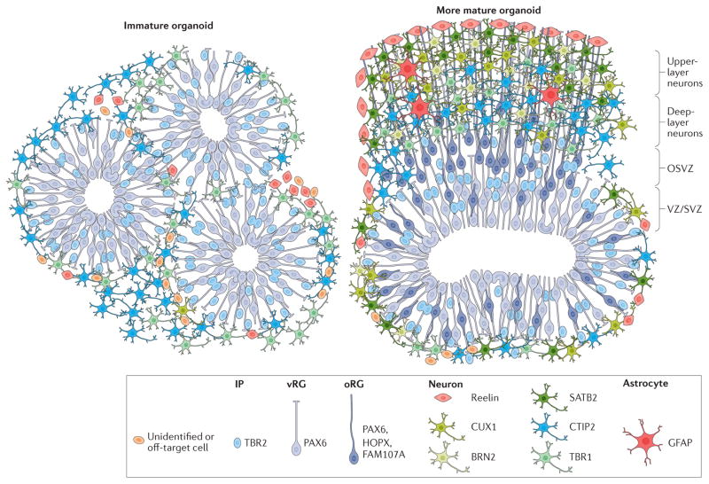
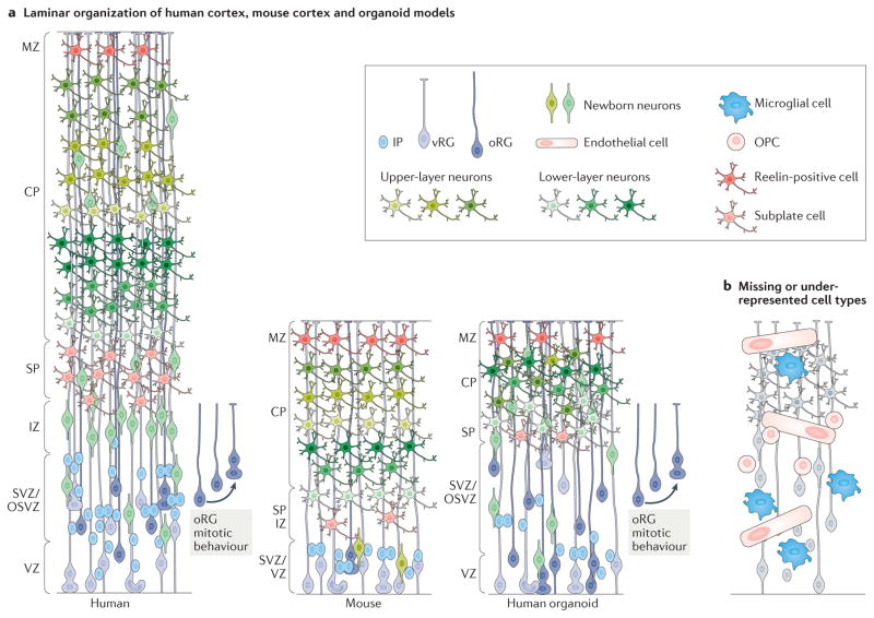
    1. Kadoshima T, et al. Self-organization of axial polarity, inside-out layer pattern, and species-specific progenitor dynamics in human ES cell-derived neocortex. Proc Natl Acad Sci USA. 2013;110:20284–20289. This study provided a detailed exploration of brain organoid tissue including the temporal and spatial organization of cell type diversity and patterning. - PMC - PubMed
    1. Jo J, et al. Midbrain-like organoids from human pluripotent stem cells contain functional dopaminergic and neuromelanin-producing neurons. Cell Stem Cell. 2016;19:248–257. - PMC - PubMed
    1. Qian X, et al. Brain-region-specific organoids using mini-bioreactors for modeling ZIKV exposure. Cell. 2016;165:1238–1254. This study used mini bioreactors to produce forebrain organoids with a well-defined OSVZ and demonstrated the presence of oRGs with defined molecular markers. Diverse neuronal cell types expressing molecular markers of all six cortical layers were observed. - PMC - PubMed

Publication types