

Review: Medial collateral ligament injuries
- PMID: 28878515
- PMCID: PMC5581380
- DOI: 10.1016/j.jor.2017.07.017
Review: Medial collateral ligament injuries
Abstract
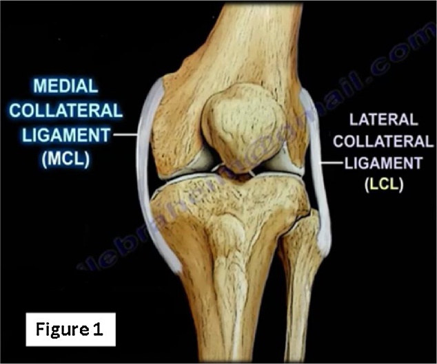
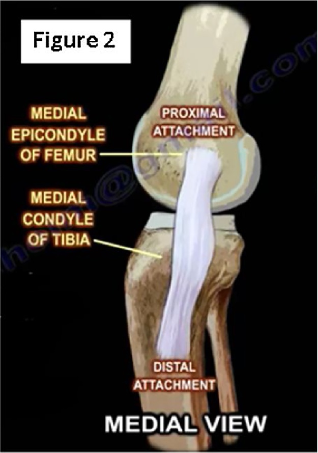
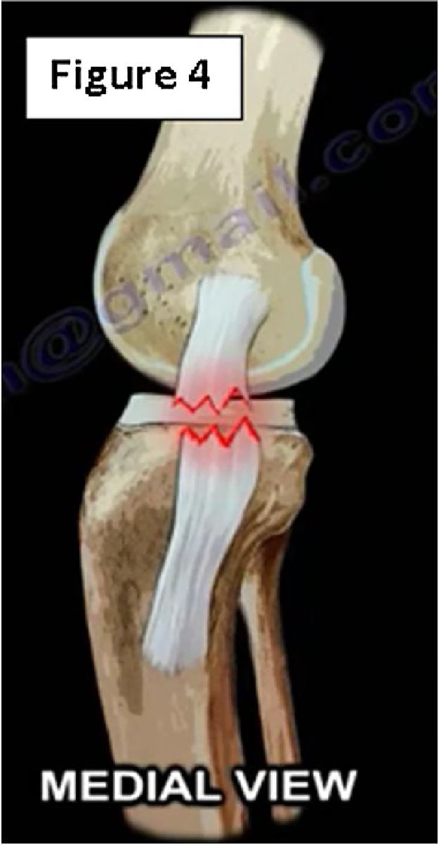
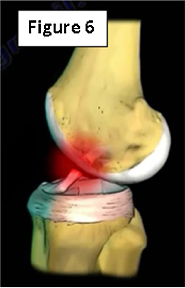
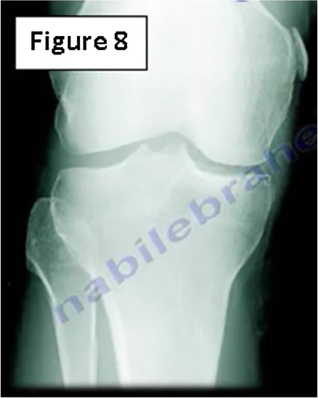
The medial collateral ligament (MCL) is a major stabilizer of the knee joint. It is the most common ligament injured in the knee, particularly in athletes, and has been reported to be torn in 7.9% of all knee injuries.2 The MCL has a complex, layered anatomy with multiple insertions and functions. Minor trauma can cause tearing of the superficial portion whereas higher energy mechanisms can disrupt both the deep and superficial layers. History and physical are often adequate, but the gold standard for diagnosis is MRI. Lesser injuries to the MCL can often be treated conservatively with early rehabilitation, but more significant tears often necessitate surgery. A thorough understanding of the MCL and associated injuries is essential for proper diagnosis and treatment.
Keywords: Anatomy; Knee; MCL; Medial collateral ligament; Pellegrini-stieda; Sports injury; Valgus stress.
Figures


References
-
- Majewski M., Susanne H., Klaus S. Epidemiology of athletic knee injuries: a 10-year study. Knee. 2006;13(3):184–188. - PubMed
-
- Warren L.F., Marshall J.L. The supporting structures and layers on the medial side of the knee. J Bone Joint Surg. 1979;61(1):56–62. - PubMed
-
- Laprade R.F. The anatomy of the medial part of the knee. J Bone Joint Surg (Am) 2007;89(9):2000. - PubMed
-
- Lind M., Jakobsen B.W., Lund B., Hansen M.S., Abdallah O., Christiansen S.E. Anatomical reconstruction of the medial collateral ligament and posteromedial corner of the knee in patients with chronic medial collateral ligament instability. Am J Sports Med. 2009;37(6):1116–1122. - PubMed
Publication types
LinkOut - more resources
Full Text Sources
Other Literature Sources
