Review
. 2017 Aug 22:10:263.
doi: 10.3389/fnmol.2017.00263. eCollection 2017.

Autophagy Dysregulation in ALS: When Protein Aggregates Get Out of Hand

Affiliations
Review

Autophagy Dysregulation in ALS: When Protein Aggregates Get Out of Hand

Nandini Ramesh et al. Front Mol Neurosci. .

Abstract

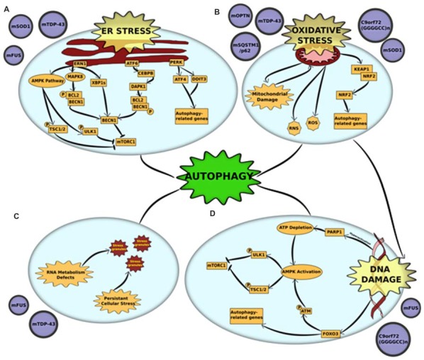
Amyotrophic lateral sclerosis (ALS) is a neurodegenerative disorder that results from the loss of upper and lower motor neurons. One of the key pathological hallmarks in diseased neurons is the mislocalization of disease-associated proteins and the formation of cytoplasmic aggregates of these proteins and their interactors due to defective protein quality control. This apparent imbalance in the cellular protein homeostasis could be a crucial factor in causing motor neuron death in the later stages of the disease in patients. Autophagy is a major protein degradation pathway that is involved in the clearance of protein aggregates and damaged organelles. Abnormalities in autophagy have been observed in numerous neurodegenerative disorders, including ALS. In this review, we discuss the contribution of autophagy dysfunction in various in vitro and in vivo models of ALS. Furthermore, we examine the crosstalk between autophagy and other cellular stresses implicated in ALS pathogenesis and the therapeutic implications of regulating autophagy in ALS.

Keywords: ALS; C9orf72; FUS; SOD1; TDP-43; autophagy; motor neuron disease; neurodegeneration.


Figures

Figure 1
Figure 1
Cross-talk between autophagy and other pathogenic mechanisms in amyotrophic lateral sclerosis (ALS) (A) endoplasmic reticulum (ER) stress and unfolded protein response (UPR) is triggered by mutations in superoxide dismutase 1 (mSOD1), TAR DNA-binding protein 43 (TDP-43) and fused in sarcoma (FUS). Accumulation of misfolded mutant proteins activates either one or all three UPR pathways—protein kinase RNA-like endoplasmic reticulum kinase (PERK), endoplasmic reticulum to nucleus signaling 1 (ERN1) and activating transcription factor 6 (ATF6). Each of the pathways stimulate autophagy through downstream activators. ERN1 activates autophagy through three pathways—(i) the AMPK pathway phosphorylates and activates two proteins—ULK1 and tuberous sclerosis protein 1/2 (TSC1/2)—both of which inhibit mammalian target of rapamycin complex 1 (mTORC1) which leads to initiation of autophagy; (ii) ERN1 activates mitogen-activated protein kinase 8 (MAPK8), which uses its kinase activity to phosphorylate B-cell lymphoma 2 (BCL2) which is in a complex with Beclin-1 (BECN1). This phosphorylation disrupts the complex and releases BECN1, which in turn activates autophagy through mTORC1 inhibition; (iii) ERN1 also acts an endoribonuclease to splice XBP1. The spliced version of XBP1 (XBP1s) promotes expression of BECN1. The UPR activator ATF6 activates the transcription factor CCAAT/enhancer-binding protein beta (CEBPB) that promotes expression of the kinase death-associated protein kinase 1 (DAPK1). DAPK1 phosphorylates BECN1 and thus breaks it from its complex with BCL2. The free BECN1 now activates autophagy through mTORC1 inhibition. The third UPR activator, PERK, activates the expression of two stress response genes—activating transcription factor 4 (ATF4) and DNA damage inducible transcript 3 (DDIT3). These genes promote the transcription of autophagy-related genes (ATGs) that activate the process. (B) Oxidative stress and mitochondrial dysfunction are complimentary mechanisms that are triggered by mutations in optineurin (mOPTN), TDP-43, SQSTM1/p62, SOD1 and C9orf72 GGGGCC expansion. Mitochondrial dysfunction is the primary source of reactive oxygen and reactive nitrogen species (ROS and RNS) that are directly implicated in activating autophagy. An alternative mechanism is through the Kelch-like ECH-associated protein 1 (KEAP1)-nuclear factor erythroid 2-related factor 2 (NRF2) pathways. Oxidative stress KEAP1-NRF2 complex is disrupted. This leaves NRF2 free to translocate to the nucleus where it induces the expression of several autophagy-related genes. Autophagy also acts to clear damaged mitochondria through an organelle-specific mechanism called mitophagy. (C) RNA metabolism defect is a major pathological outcome of mutations in RNA-binding proteins, especially FUS and TDP-43. These proteins are major components of stress granules and mutations in low complexity domains of these proteins alter the phase of stress granules to give them a complex liquid state. This alters the dynamics of stress granules, in that, they are not cleared and potentially act as primers for protein aggregates. Autophagy is a crucial mechanism involved in the clearance of stress granules under normal conditions and dysfunction in autophagy could be linked to altered stress granule dynamics. (D) DNA damage, either caused as a direct result of pathogenic mutation or triggered by oxidative stress, is a common pathological feature of mutations in FUS and C9orf72 GGGGCC expansion. DNA damage activates autophagy through activation of AMPK pathway. The AMPK pathway phosphorylates and activates two proteins, ULK1 and TSC1/2, both of which independently act as mTORC1 inhibitors. DNA damage activates the AMPK pathways through: (i) Hyperactivation of PARP1, which leads to a depletion of NAD+ reserves in the cell. This, in turn, depletes ATP and thus activates AMPK; or (ii) FOXOa3 dissociation from DNA that allows it to interact with ATM. Activation of ATM by phosphorylation leads to downstream activation of AMPK pathway. Alternatively, FOXOa3 also acts as a transcription factor that regulates expression of autophagy-related genes.

References

    1. Abe K., Itoyama Y., Sobue G., Tsuji S., Aoki M., Doyu M., et al. . (2014). Confirmatory double-blind, parallel-group, placebo-controlled study of efficacy and safety of edaravone (MCI-186) in amyotrophic lateral sclerosis patients. Amyotroph. Lateral Scler. Frontotemporal Degener. 15, 610–617. 10.3109/21678421.2014.959024 - DOI - PMC - PubMed
    1. Abe K., Yuki S., Kogure K. (1988). Strong attenuation of ischemic and postischemic brain edema in rats by a novel free radical scavenger. Stroke 19, 480–485. 10.1161/01.str.19.4.480 - DOI - PubMed
    1. Almeida S., Gascon E., Tran H., Chou H. J., Gendron T. F., Degroot S., et al. . (2013). Modeling key pathological features of frontotemporal dementia with C9ORF72 repeat expansion in iPSC-derived human neurons. Acta Neuropathol. 126, 385–399. 10.1007/s00401-013-1149-y - DOI - PMC - PubMed
    1. Al-Sarraj S., King A., Troakes C., Smith B., Maekawa S., Bodi I., et al. . (2011). p62 positive, TDP-43 negative, neuronal cytoplasmic and intranuclear inclusions in the cerebellum and hippocampus define the pathology of C9orf72-linked FTLD and MND/ALS. Acta Neuropathol. 122, 691–702. 10.1007/s00401-011-0911-2 - DOI - PubMed
    1. Arai T., Hasegawa M., Akiyama H., Ikeda K., Nonaka T., Mori H., et al. . (2006). TDP-43 is a component of ubiquitin-positive tau-negative inclusions in frontotemporal lobar degeneration and amyotrophic lateral sclerosis. Biochem. Biophys. Res. Commun. 351, 602–611. 10.1016/j.bbrc.2006.10.093 - DOI - PubMed