. 2017 Sep 4:8:14.
doi: 10.1186/s13227-017-0077-7. eCollection 2017.

PaxA, but not PaxC, is required for cnidocyte development in the sea anemone Nematostella vectensis

Affiliations

PaxA, but not PaxC, is required for cnidocyte development in the sea anemone Nematostella vectensis

Leslie S Babonis et al. Evodevo. .

Abstract

Background: Pax genes are a family of conserved transcription factors that regulate many aspects of developmental morphogenesis, notably the development of ectodermal sensory structures including eyes. Nematostella vectensis, the starlet sea anemone, has numerous Pax orthologs, many of which are expressed early during embryogenesis. The function of Pax genes in this eyeless cnidarian is unknown.

Results: Here, we show that PaxA, but not PaxC, plays a critical role in the development of cnidocytes in N. vectensis. Knockdown of PaxA results in a loss of developing cnidocytes and downregulation of numerous cnidocyte-specific genes, including a variant of the transcription factor Mef2. We also demonstrate that the co-expression of Mef2 in a subset of the PaxA-expressing cells is associated with the development with a second lineage of cnidocytes and show that knockdown of the neural progenitor gene SoxB2 results in downregulation of expression of PaxA, Mef2, and several cnidocyte-specific genes. Because PaxA is not co-expressed with SoxB2 at any time during cnidocyte development, we propose a simple model for cnidogenesis whereby a SoxB2-expressing progenitor cell population undergoes division to give rise to PaxA-expressing cnidocytes, some of which also express Mef2.

Discussion: The role of PaxA in cnidocyte development among hydrozoans has not been studied, but the conserved role of SoxB2 in regulating the fate of a progenitor cell that gives rise to neurons and cnidocytes in Nematostella and Hydractinia echinata suggests that this SoxB2/PaxA pathway may well be conserved across cnidarians.

Keywords: Cell differentiation; Evolution; Gene regulatory network; Mef2; Nematocyte; Novelty; PaxA; PaxC; SoxB2.


Figures

Fig. 1
Fig. 1
Type and distribution of cnidocytes in N. vectensis. a Microbasic p-mastigophores have a thick shaft which ends in a distinctive v-shaped notch (arrow). b Basitrichous isorhizas are variable in size (compare left and right DIC images) and have a thin shaft lacking a v-shaped notch and a tubule which is visible through the capsule (arrows). c Spirocytes can be identified by the regularly spaced coiled tubule and the lack of a visible capsule (arrow); a basitrichous isorhiza is shown for comparison (arrowhead). d, e The mesenterial filaments and pharynx are populated largely by microbasic p-mastigophores and large basitrichous isorhizas. f The body wall is populated by small basitrichs, and g the tentacle tips are populated by large basitrichous isorhizas (arrowheads in c, g) and spirocytes (arrows in c, g). Only nematocytes (basitrichous isorhizas and microbasic p-mastigophores) can be labeled using DAPI, indicated by yellow color in the cartoons (a, b). Scale bars in ac represent 5 μm. All images are DIC micrographs
Fig. 2
Fig. 2
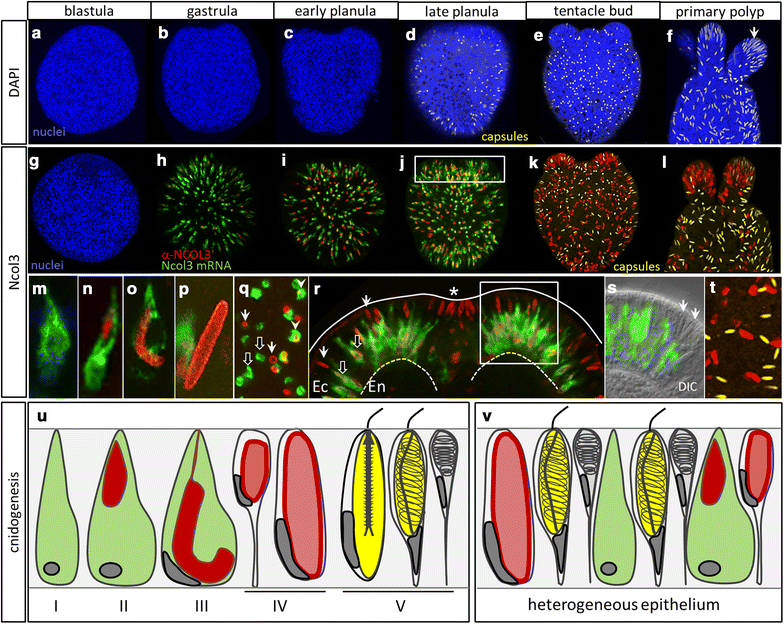
Stages of cnidogenesis in N. vectensis. af Mature nematocyst capsules (yellow, DAPI) are first detected in the ectoderm in the late planula stage and are dense in the tentacle tips at the polyp stage (f, arrow). gl Expression of Ncol3 mRNA (green) and NCOL3 protein (red) at the gastrula stage (h). New cnidocytes continue to develop in the embryonic ectoderm at all stages, even when mature cnidocytes (yellow, DAPI) are present in this tissue (k, l). ms Multiple stages of cnidocytes are present in the ectoderm at the same time. Early-stage cnidocytes express Ncol3 mRNA only (m); later, many developing cnidocytes express mRNA and translated protein at the same time (n, o). Late-stage cnidocytes express protein only (P) and appear like hollow cylinders (arrows, q). r In the late planula, developing cnidocytes are dense in the ectoderm (Ec) adjacent to the blastopore (asterisk). Early-stage cnidocytes (black arrows) appear shifted toward the mesoglea side of the ectoderm (dotted line), and late-stage cnidocytes (white arrows) appear shifted toward the apical side of the ectoderm (solid line). (r corresponds to the boxed region in j.) s Cells expressing Ncol3 mRNA only continue to develop in the embryonic ectoderm, even in the presence of mature cnidocytes (arrows, s). (s corresponds to the boxed region in r). t NCOL3 protein (red) and DAPI (yellow) do not co-localize at any stage of development. u Cartoon summary of cnidocyte development as observed through Ncol3 and DAPI labeling. Colors correspond to the labeling in at. Nuclei are blue, DAPI. The blastopore/oral pole is oriented to the top unless otherwise indicated by asterisk for this and all subsequent figures. Images in al are 3D renderings from confocal z-stacks; images mt are single optical sections
Fig. 3
Fig. 3
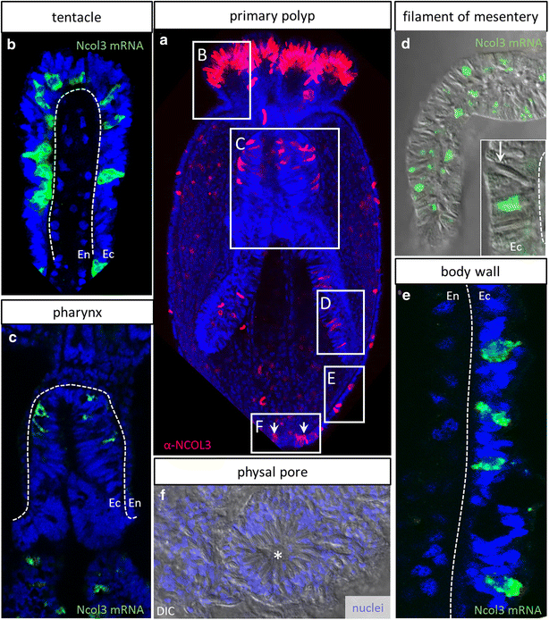
Distribution and development of new cnidocytes in tissues of the polyp. a Four-tentacle primary polyp illustrating the development of new cnidocytes (NCOL3 protein, red) in the ectoderm of multiple tissues. be Individual tissues from eight-tentacle-stage polyps showing expression of Ncol3 mRNA (green) in the ectodermally derived tissue (Ec) only (dotted line indicates mesoglea; En—endodermally derived tissue). The arrow in d points to a mature cnidocyte in the cnidoglandular (ectodermal) portion of the mesenterial filament. f Aggregation of mature cnidocytes encircling an ephemeral opening (asterisk) at the aboral pole of the adult polyp. The arrows in a correspond to new cnidocytes developing in the region highlighted in f. a 3D rendering from a confocal z-stack; b, c, e individual optical sections; d, f DIC micrographs
Fig. 4
Fig. 4
Minicollagen mRNAs are co-expressed, but the onset of NCOL protein localization is variable. ad Ncol3 and Ncol4 mRNAs are co-expressed in all developing cnidocytes at the tentacle bud stage. eh Ncol3 and Ncol1 mRNAs are also largely co-expressed, though cnidocytes that appear to be expressing Ncol3 exclusively can be seen in the ectoderm (arrows f, g). il Ncol4 and Ncol1 mRNAs are co-expressed in all cnidocytes. mp NCOL3 and NCOL4 proteins largely co-localize, though early-stage capsules may express only NCOL3 (arrows n, o) and late-stage capsules may express only NCOL4 (arrowheads o, p). qt NCOL1 and NCOL4 proteins also co-localize in all developing cnidocytes, though some late-stage capsules may express only NCOL4 (arrows r, t). u In gastrula-stage embryos, cnidocytes comprise approximately 10% of the cells, independent of which antibody is used (NCOL1: 9.39 ± 0.59, NCOL3: 11.01 ± 0.81, NCOL4: 9.11 ± 0.68; mean ± standard error). N = number of embryos analyzed. (V-AG) Co-labeling with mRNA probes and antibodies for the corresponding protein confirms that transcription is downregulated before cnidocyst capsules are fully polymerized as proteins can be detected in cells that no longer express mRNA for each antibody/probe combination (arrows w, aa, ae). ahak Co-labeling of Ncol3 mRNA with α-NCOL4 also reveals many late-stage cnidocytes labeled with α-NCOL4 only (arrows aj). Main images are 3D renderings from confocal z-stacks, high-magnification panels are single optical sections
Fig. 5
Fig. 5
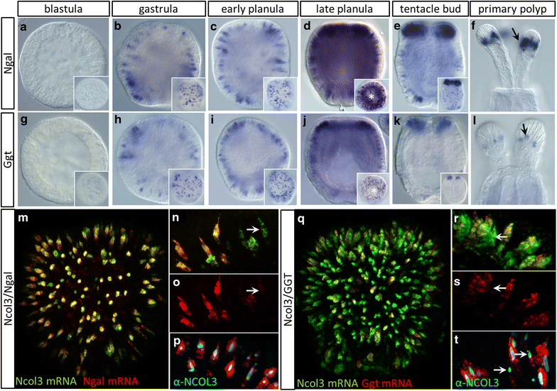
Characterization of two novel markers for differentiated cnidocytes. Expression of af nematogalectin (Ngal) mRNA and gl γ-glutamyl transpeptidase (Ggt) mRNA during development. Onset of expression begins at/before the gastrula stage in scattered cells of the ectoderm (surface plane, insets) for both Ngal (b) and Ggt (h). Ngal is expressed in the body wall ectoderm throughout development (be, insets), and expression becomes dense in the developing tentacle buds (e) and tentacle tips of the polyp (f, arrow). Ggt is also expressed in the embryonic ectoderm (hj) and presumptive tentacle buds but is absent from the body wall at later stages (k, inset) and is expressed in only few cells of the tentacle tips (l, arrow). m Ngal mRNA is co-expressed with Ncol3 mRNA in most developing cnidocytes in early-planula-stage embryos. n, o Few cells exhibit Ncol3 mRNA expression without Ngal (arrows), but p Ngal mRNA and NCOL3 protein are perfectly co-expressed. qs Ggt mRNA is co-expressed with Ncol3 mRNA in many but not all developing cnidocytes. (Arrows in rs indicate cells expressing Ncol3 only.) t Co-labeling of NCOL3 protein and Ggt mRNA confirms that some cnidocytes express Ncol3 without Ggt (arrows). Images in al are DIC micrographs, images m and q are 3D renderings from confocal z-stacks, and images np and rt are individual optical sections
Fig. 6
Fig. 6
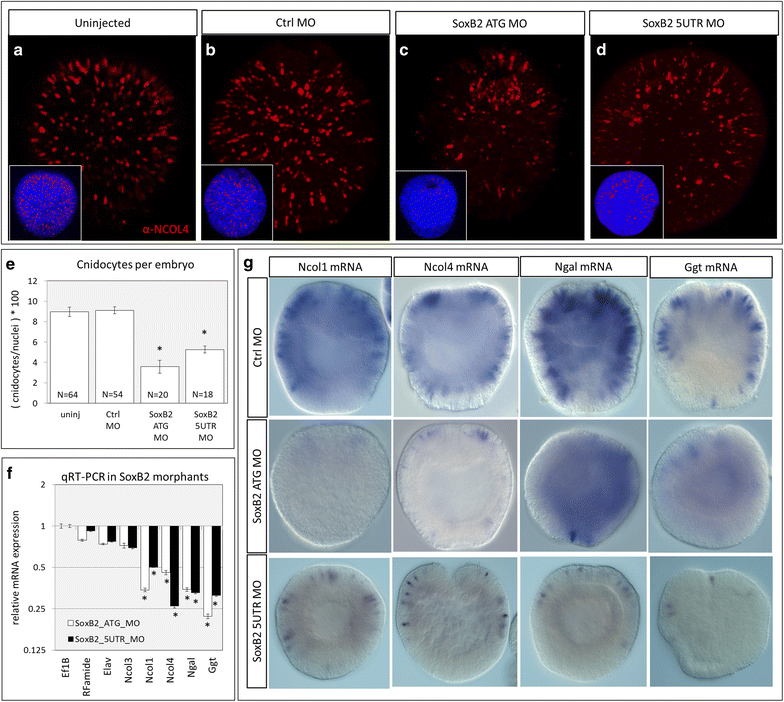
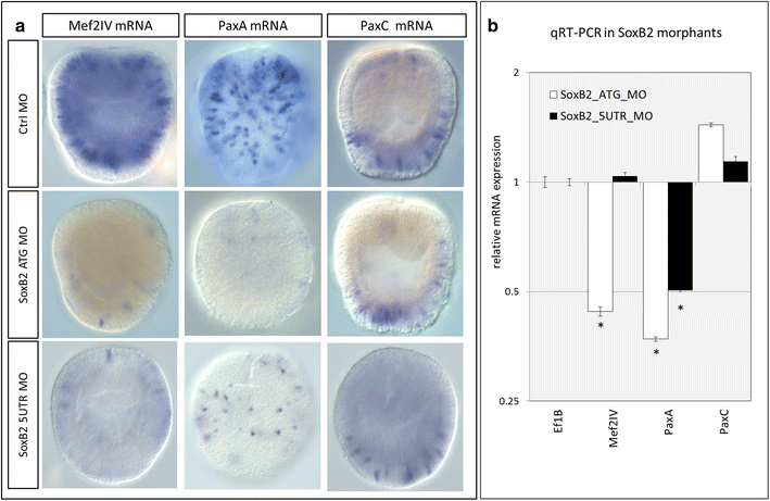
Identification of putative cnidocyte-specific transcription factors. ad Cnidocyte abundance was characterized in gastrula-stage embryos labeled with α-NCOL4 antibody in a unmanipulated embryos or following microinjection of b standard control morpholino (Ctrl MO), c SoxB2 ATG MO, or d SoxB2 5UTR MO. Nuclei were labeled with DAPI (insets), and cnidocyte abundance was measured relative to the total number of labeled nuclei in each embryo. e Cnidocytes comprise approximately 9% of the cells of the ectoderm in uninjected and control MO-injected embryos and only 4% of the cells in SoxB2 MO embryos (uninjected: 8.98 ± 0.45, control MO: 9.11 ± 0.33, SoxB2 ATG MO: 3.82 ± 1.84, SoxB2 5UTR MO: 5.27 ± 0.34; mean ± standard error). Asterisk indicates significant difference from control MO-injected embryos. f Relative mRNA expression of target genes assayed by qRT-PCR following microinjection of SoxB2 MO. The expression of housekeeping gene EF1B in SoxB2 morphants relative to embryos injected with control MO was set to 1.0, and all other expression values are reported relative to this. The y-axis is presented in log scale. Mean ± standard error. Asterisk indicates significant difference from Ef1B. g In situ hybridization of cnidocyte-specific genes in control MO-injected and SoxB2 MO-injected embryos validating the results of qRT-PCR analysis. Images in g are DIC micrographs
Fig. 7
Fig. 7
Mef2IV and PaxA, but not PaxC, are downstream of SoxB2. a Spatial expression of Mef2IV, PaxA, and PaxC in gastrula-stage embryos injected with control and SoxB2 morpholinos. b qRT-PCR in SoxB2 morphants demonstrates knockdown of Mef2IV (5UTR MO only) and PaxA, but not PaxC. Asterisk indicates significant difference from Ef1B. Images in a are DIC micrographs
Fig. 8
Fig. 8
Co-expression of Mef2IV and PaxA, not PaxC, in developing cnidocytes. ac Mef2IV mRNA co-localizes with Ncol3 mRNA in only ~25% of the developing cnidocytes. (Cells co-expressing both markers are indicated by arrows, and cells expressing one marker only are indicated by circles in panels aj). df PaxA mRNA co-localizes with Ncol3 mRNA in ~50% of the developing cnidocytes. gj PaxA mRNA is co-expressed in Mef2IV-expressing cells, but several cells express PaxA without Mef2IV. Co-localization of k, l Mef2IV mRNA and m, n PaxA mRNA with NCOL3 protein. or Co-labeling of PaxA and Mef2IV mRNA with α-NCOL3 indicates there are cells expressing all three markers at the same time (arrows), cells expressing PaxA and NCOL3 without Mef2IV (dotted circles), and cells expressing NCOL3 protein without either PaxA or Mef2IV (arrowhead). sv PaxC is not co-expressed with Ncol3 or wz PaxA. aaad PaxA and SoxB2 are not co-expressed. Insets and main images are 3D reconstructions from confocal z-stacks; high-magnification images are individual optical sections
Fig. 9
Fig. 9
Knockdown of PaxA results in loss of cnidocytes. a The PaxA sp MO recognizes the I1E2 boundary which should cause skipping of exon 2, but the PaxA transcript could only be amplified from embryos injected with control morpholino (Ctrl MO). b The PaxC sp MO blocks the E1I1 boundary causing retention of intron 1. Both WT and morphant transcripts were amplified from PaxC MO-injected embryos. cg Developing cnidocytes labeled with α-NCOL4 antibody in embryos injected with c control morpholino, d PaxA splice-blocking morpholino, e PaxA translation-blocking morpholino, f PaxC splice-blocking morpholino, and g PaxC translation-blocking morpholino. h Quantitative analysis of cnidocyte abundance in early-planula-stage embryos. Control morpholino-injected embryos exhibited wild-type abundance of cnidocytes, whereas PaxA sp morphants and PaxA tr morphants had significantly fewer (control MO: 9.11 ± 0.39, PaxA sp: 3.65 ± 0.63, PaxA tr: 4.90 ± 0.66; mean ± standard error). Both PaxC sp morphants and PaxC tr morphants exhibited an increase in cnidocyte abundance, albeit with a small effect size (PaxC sp: 10.58 ± 0.68, PaxC tr: 12.44 ± 0.47). i PaxA mRNA partially rescues SoxB2 ATG morphant phenotype but is not sufficient to induce ectopic cnidocytes. Asterisk indicates significant difference from Ctrl MO. Dagger indicates significant difference from SoxB2 ATG MO
Fig. 10
Fig. 10
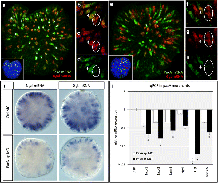
Knockdown of PaxA results in downregulation of cnidocyte-specific genes. ah PaxA is largely co-expressed with ad Ngal and eh Ggt. Arrows indicate cells expressing both markers, and dotted circles indicate cells expressing Ngal or Ggt only. i Knockdown of PaxA results in a decrease in the number of cells expressing Ngal and Ggt (relative to control morpholino) and j downregulation of other cnidocyte-specific genes, relative to embryos injected with control MO. Asterisk indicates significant difference relative to Ef1B expression. Main images a, e are 3D renderings of confocal z-stacks; high-magnification images are single optical sections. Images in i are DIC micrographs
Fig. 11
Fig. 11
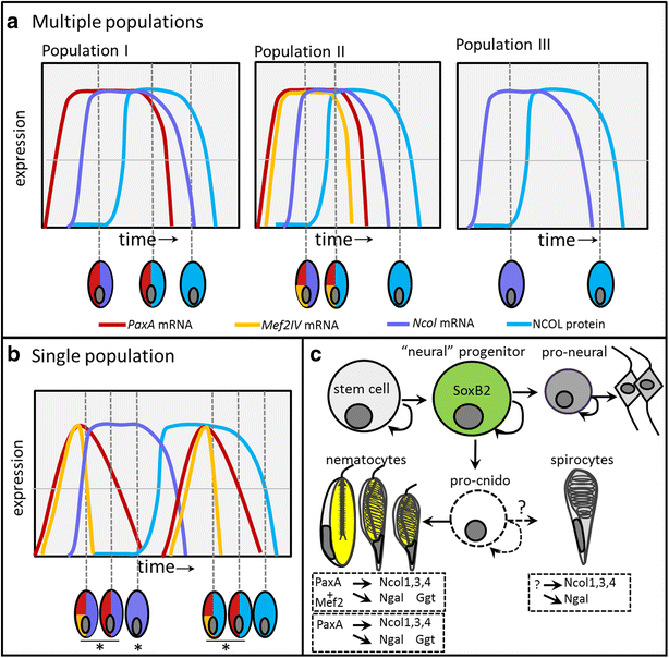
Cnidogenesis in N. vectensis is temporally and spatially dynamic. a Hypothesis 1: Cnidocytes develop from multiple independent populations of cells all present in the same epithelium at the same time. b Hypothesis 2: There is a single population of developing cnidocytes and all combinations of gene expression presented here result from temporal shifts in gene expression. In both cases, Ncol mRNA (purple) must be expressed before NCOL protein is detectable (blue) and, when expressed, PaxA (red) and Mef2IV (yellow) must be upregulated before Ncol mRNA. Horizontal lines in a, b represent an arbitrary threshold of detectable expression. c SoxB2-expressing progenitor cells give rise to both neurons and cnidocytes in N. vectensis. Post-mitotic neurons differentiate from cells expressing pro-neural genes, though the existence of “pro-cnido” genes (expressed in a progenitor that gives rise only to cnidocytes) is still only hypothesized (indicated by dotted lines). Three populations of cnidocytes may be present. Two populations differ only in the expression of Mef2IV and are hypothesized to become either basitrichous isorhizas and microbasic p-mastigophores or the two distinct size classes of basitrichous isorhizas. A third population expresses Ncol1, Ncol3, Ncol4, and Ngal, but the transcription factor that activates expression of these cnidocyte-specific genes is unknown. We hypothesize that this population of cnidocytes becomes spirocytes

References

    1. David CN, Ozbek S, Adamczyk P, Meier S, Pauly B, Chapman J, Hwang JS, Gojobori T, Holstein TW. Evolution of complex structures: minicollagens shape the cnidarian nematocyst. Trends Genet. 2008;24:431–438. doi: 10.1016/j.tig.2008.07.001. - DOI - PubMed
    1. Galliot B, Quiquand M. A two-step process in the emergence of neurogenesis. Eur J Neurosci. 2011;34:847–862. doi: 10.1111/j.1460-9568.2011.07829.x. - DOI - PubMed
    1. Galliot B, Quiquand M, Ghila L, de Rosa R, Miljkovic-Licina M, Chera S. Origins of neurogenesis, a cnidarian view. Dev Biol. 2009;332:2–24. doi: 10.1016/j.ydbio.2009.05.563. - DOI - PubMed
    1. Kass-Simon G, Scappaticci JAA. The behavioral and developmental physiology of nematocysts. Can J Zool. 2002;80:1772–1794. doi: 10.1139/z02-135. - DOI
    1. Kurz EM, Holstein TW, Petri BM, Engel J, David CN. Mini-collagens in Hydra nematocytes. J Cell Biol. 1991;115:1159–1169. doi: 10.1083/jcb.115.4.1159. - DOI - PMC - PubMed

LinkOut - more resources