

Progressive medial temporal lobe atrophy during preclinical Alzheimer's disease
- PMID: 28879085
- PMCID: PMC5577409
- DOI: 10.1016/j.nicl.2017.08.022
Progressive medial temporal lobe atrophy during preclinical Alzheimer's disease
Abstract
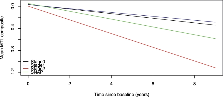
This study examined whether longitudinal MRI trajectories in medial temporal lobe (MTL) brain regions differed among groups of cognitively normal individuals defined by their cerebrospinal fluid (CSF) levels when they were first enrolled (N = 207; mean clinical follow-up = 13.3 years (max = 20 years), mean MRI follow-up = 2.4 years (max = 8 years)). We first compared atrophy rates among groups defined by CSF amyloid and phosphorylated-tau (p-tau) vs. CSF amyloid and total tau (t-tau). We also examined whether, in the presence of amyloid or tau/p-tau, the atrophy rates differed based on whether the subjects ultimately progressed to a diagnosis of mild cognitive impairment (MCI), as well as whether apolipoprotein ε4 (Apoε4) status had an impact on the longitudinal MRI trajectories. The primary finding was that when the groups were defined using CSF amyloid and p-tau, individuals with low levels of CSF amyloid and high levels of CSF p-tau (referred to as Stage 2) showed a significantly greater rate of atrophy in a composite measure of MTL volumes compared to groups defined by evidence of abnormal CSF levels in only one of the brain proteins (but not both), or no evidence of CSF abnormality. In contrast, there were no differences in rate of MTL atrophy when the groups were defined by levels of CSF amyloid and t-tau (instead of p-tau). Additionally, the rate of MTL atrophy did not differ between subjects who progressed to MCI at follow-up vs. those who remained cognitively normal when CSF levels of amyloid, t-tau, or p-tau were covaried. Lastly, the presence of an APOE ε4 genotype did not modulate the degree of MTL atrophy once baseline levels of CSF amyloid, p-tau or t-tau were accounted for. These results suggest that abnormal levels of CSF amyloid and CSF p-tau (but not t-tau) maximize the likelihood of observing significant MTL atrophy over time among individuals with normal cognition at baseline, and emphasize the importance of differentiating biomarkers that primarily reflect neurofibrillary tangle pathology (CSF p-tau) compared with biomarkers of neuronal injury (CSF t-tau).
Keywords: Amyloid; Cerebrospinal fluid; Magnetic resonance imaging; Phosphorylated tau; Preclinical AD.
Figures
References
-
- Albert DeKosky S.T., Dickson D., Dubois B., Feldman H.H., Fox N.C., Gamst A., Holtzman D.M., Jagust W.J., Petersen R.C., Snyder P.J., Carrillo M.C., Thies B., Phelps C.H. The diagnosis of mild cognitive impairment due to Alzheimer's disease: recommendations from the National Institute on Aging-Alzheimer's Association workgroups on diagnostic guidelines for Alzheimer's disease. Alzheimers Dement. 2011;7:270–279. - PMC - PubMed
-
- Beg M.F., Miller M.I., Trouve A., Younes L. Computing metrics via geodesics on flows of diffeomorphisms. Int. J. Comput. Vis. 2005;61:139–157.
-
- Buerger K., Ewers M., Pirttila T., Zinkowski R., Alafuzoff I., Teipel S.J., DeBernardis J., Kerkman D., McCulloch C., Soininen H., Hampel H. CSF phosphorylated tau protein correlates with neocortical neurofibrillary pathology in Alzheimer's disease. Brain. 2006;129:3035–3041. - PubMed
-
- Corder E.H., Saunders A.M., Risch N.J., Strittmatter W.J., Schmechel D.E., Gaskell P.C., Jr., Rimmler J.B., Locke P.A., Conneally P.M., Schmader K.E. Protective effect of apolipoprotein E type 2 allele for late onset Alzheimer disease. Nat. Genet. 1994;7:180–184. - PubMed
MeSH terms
Substances
Grants and funding
LinkOut - more resources
Full Text Sources
Other Literature Sources
Medical
Miscellaneous
