

Faciobrachial dystonic seizures result from fronto-temporo-basalganglial network involvement
- PMID: 28879090
- PMCID: PMC5573798
- DOI: 10.1016/j.ebcr.2017.06.001
Faciobrachial dystonic seizures result from fronto-temporo-basalganglial network involvement
Abstract
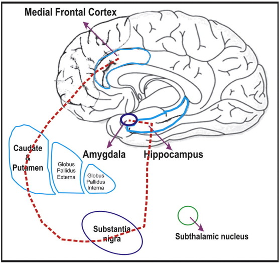
•Faciobrachial dystonic seizures (FBDS) are caused by autoantibodies to leucine-rich glioma-inactivated1 proteins, a component of the voltage-gated potassium channel complex (VGKC-complex) and precede the clinical presentation of limbic encephalitis.•The exact pathophysiology of FBDS is not known and whether they are seizures or movement disorder is still debated.•We suggest the fronto-temporo-basal ganglia network involving the medial frontal and temporal regions along with the corpus striatum and substantia nigra being responsible for the clinical phenomenon of FBDS.•The varied clinical, electrical and imaging features of FBDS in our cases and in the literature are best explained by involvement of this network.•Entrainment from any part of this network will result in similar clinical expression of FBDS, whereas other electro-clinical associations and duration depends on the extent of involvement of the network.
Keywords: Basalganglial hypermetabolism; Faciobrachial dystonic seizures; Fronto–temporo–basalganglial network; LGI1antibody; Network hypothesis; VGKC antibody encephalitis.
Figures

References
-
- Irani S.R., Buckley C., Vincent A. Immunotherapy responsive seizure-like episodes with potassium channel antibodies. Neurology. 2008;71:1647–1648. - PubMed
-
- Irani S.R., Michell A.W., Lang B. Faciobrachial dystonic seizures precede Lgi1 antibody limbic encephalitis. Ann Neurol. 2011;69:892–900. - PubMed
-
- Irani S.R., Stagg C.J., Schott J.M. Faciobrachial dystonic seizures: the influence of immunotherapy on seizure control and prevention of cognitive impairment in a broadening phenotype. Brain. 2013;136:3151–3162. - PubMed
-
- Striano P. Faciobrachial dystonic attacks: seizures or movement disorder? Ann Neurol. 2011;70:179–180. [author reply 180] - PubMed
Publication types
LinkOut - more resources
Full Text Sources
Other Literature Sources
