. 2017 Sep 1;3(9):e1600743.
doi: 10.1126/sciadv.1600743. eCollection 2017 Sep.

Polarization recovery through scattering media

Affiliations

Polarization recovery through scattering media

Hilton B de Aguiar et al. Sci Adv. .

Abstract

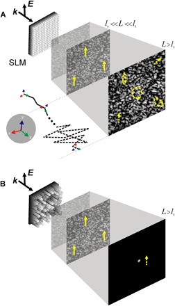
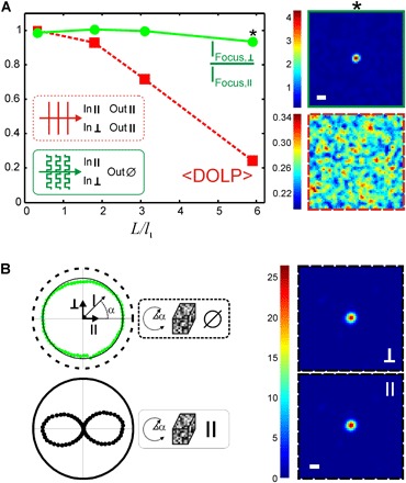
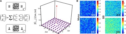
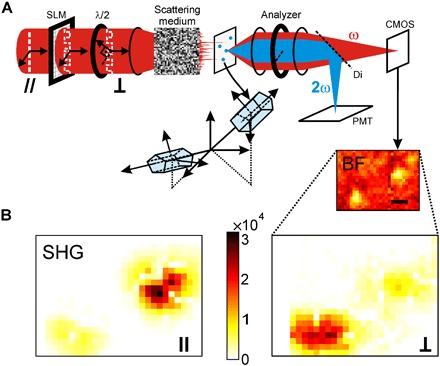
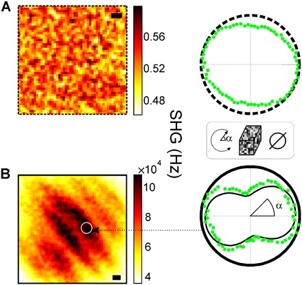
The control and use of light polarization in optical sciences and engineering are widespread. Despite remarkable developments in polarization-resolved imaging for life sciences, their transposition to strongly scattering media is currently not possible, because of the inherent depolarization effects arising from multiple scattering. We show an unprecedented phenomenon that opens new possibilities for polarization-resolved microscopy in strongly scattering media: polarization recovery via broadband wavefront shaping. We demonstrate focusing and recovery of the original injected polarization state without using any polarizing optics at the detection. To enable molecular-level structural imaging, an arbitrary rotation of the input polarization does not degrade the quality of the focus. We further exploit the robustness of polarization recovery for structural imaging of biological tissues through scattering media. We retrieve molecular-level organization information of collagen fibers by polarization-resolved second harmonic generation, a topic of wide interest for diagnosis in biomedical optics. Ultimately, the observation of this new phenomenon paves the way for extending current polarization-based methods to strongly scattering environments.


Figures

Fig. 1
Fig. 1. Illustration of polarization state scrambling during propagation in a scattering medium and principle of polarization recovery via wavefront shaping.
When light propagates in a scattering medium, the wavefront is rapidly deformed into a speckle pattern (at depths comparable to the scattering mean free path ls), but polarization scrambling occurs on a different length scale. In the forward scattering regime (as typically found in biological tissues), when the thickness L is smaller than the transport mean free path lt, forward scattering events mainly conserves the initial polarization state. (A) In the diffusive regime L > lt for an unshaped wavefront, the polarization of the speckle gradually scrambles and lacks any resemblance with the input one. The bottom panel illustrates how, during propagation, each forward scattering conserves polarization (continuous line) and how polarization is mixed when entering the diffusive regime (dashed line). (B) In contrast, we observe that an optimal wavefront shaped by a spatial light modulator (SLM) not only is able to refocus light but also recovers the original polarization state even without any polarizing optics in the detection, under broadband source illumination.
Fig. 2
Fig. 2. Experimental quantification of polarization recovery.
(A) Left: Averaged DOLP of the speckle (<DOLP>) (red squares) and refocus intensity ratio after wavefront shaping (I/I; corresponding to the same wavefront but rotated input polarization) (green circles), as a function of optical depths L/lt. Polarization scrambling of the speckle occurs for lengths of the order of lt. Nevertheless, at depths of several lt, after shaping, the refocus intensity survives a change of 90° of the input polarization. Right: Speckle image after (top) and before (bottom) the wavefront shaping procedure. The scattering media are made of 5-μm-diameter polystyrene beads. The asterisk symbol (*) in the box refers to the images shown on the right. (B) Similar experiments performed using 1-mm-thick opaque acute brain slice coronal cross section as a scattering medium. Top left: Nonanalyzed refocus intensity (green circles) upon rotation of the excitation field. Right: Images show the refocus at two input polarization states (⊥ and ∥) with the same intensity scale. Bottom left: The refocus polarization state purity is evaluated by placing an analyzer and observing an extinction (black circles). Scale bars, 1 μm.
Fig. 3
Fig. 3. Experimental quantification of vectorial transmission matrix correlations and the origin of the polarization recovery.
(A) Cross-correlation of vectorial transmission matrix elements with polarization combinations xx and yy for L/lt ≈ 6. The peak confirms strong correlation between the matrix elements, thus explaining the resilience of the refocus to a polarization state change. (B) Images of the output speckle parallel (xx) and perpendicular (yx) to the input polarization state for a monochromatic (Mono) source (left panel) and a broadband (BB) source (right panel) for L/lt ≈ 6.
Fig. 4
Fig. 4. Demonstration of structural imaging through scattering media–exploiting transmission matrix correlations.
(A) Simplified experimental layout used in the experiments. Ultrashort pulse wavefronts are shaped by an SLM and focused on the scattering medium. The speckle transmitted by the scattering medium excites the nonlinear sources (nanoKTP) placed at a plane further imaged on a complementary metal-oxide semiconductor (CMOS) camera and a photomultiplier tube (PMT). (B) Demonstration of structural imaging by polarization-resolved SHG. In the first step, the vectorial transmission matrix elements tmnxx are acquired and used to raster-scan the refocus, thus generating the SHG images (left panel). The bottom-right inset in (A) shows the bright-field (BF) image at the same region of interest (ROI), where two particles can be seen. In the second step, only the excitation polarization is rotated, and a second scan is taken (5× rescaled) (right panel) using the very same tmnxx elements. Scale bar, 1 μm.
Fig. 5
Fig. 5. Applications of transmission matrix correlations for biological specimen SHG structural imaging.
(A) Raster-scanning rat tail collagen tendon placed after a thin diffuser, with an unshaped wavefront, leads to a featureless SHG image (left panel). Furthermore, the SHG intensity response upon turning the input polarization (right panel) is almost isotropic, an outcome that is not representative of the molecular structure of collagen. (B) Raster-scanning using the memory effect, from a known transmission matrix, generates morphological features reminiscent of collagen fibers (left panel). By refocusing on a specific fiber, we recorded the intensity response of the SHG signal upon turning the input polarization angle (right panel). The continuous line (black) is a fit to the data (green circles) from which we retrieve the fiber-scale nonlinear susceptibility values of collagen. The retrieved nonlinear susceptibilities reveal the molecular order of the fibers and are in agreement with previous observations. In the polar plots, the radial direction represents SHG intensity. Scale bar, 1 μm.

References

    1. Brasselet S., Polarization-resolved nonlinear microscopy: Application to structural molecular and biological imaging. Adv. Opt. Photonics 3, 205–271 (2011).
    1. Bicout D., Brosseau C., Martinez A. S., Schmitt J. M., Depolarization of multiply scattered waves by spherical diffusers: Influence of the size parameter. Phys. Rev. E 49, 1767–1770 (1994). - PubMed
    1. Xu M., Alfano R. R., Random walk of polarized light in turbid media. Phys. Rev. Lett. 95, 213901 (2005). - PubMed
    1. Mosk A. P., Lagendijk A., Lerosey G., Fink M., Controlling waves in space and time for imaging and focusing in complex media. Nat. Photonics 6, 283–292 (2012).
    1. Katz O., Small E., Bromberg Y., Silberberg Y., Focusing and compression of ultrashort pulses through scattering media. Nat. Photonics 5, 372–377 (2011).

Publication types