Review
. 2017 Sep;19 Suppl 1(Suppl 1):124-136.
doi: 10.1111/dom.13031.

Signals in the pancreatic islet microenvironment influence β-cell proliferation

Affiliations
Review

Signals in the pancreatic islet microenvironment influence β-cell proliferation

Kristie I Aamodt et al. Diabetes Obes Metab. 2017 Sep.

Abstract

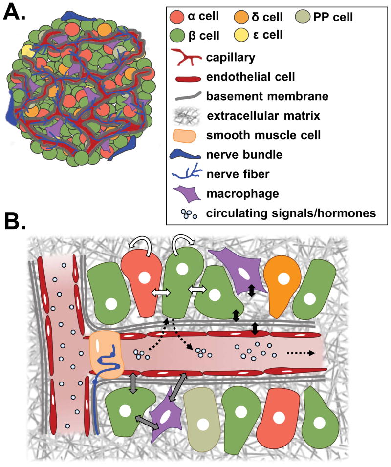
The progressive loss of pancreatic β-cell mass that occurs in both type 1 and type 2 diabetes is a primary factor driving efforts to identify strategies for effectively increasing, enhancing or restoring β-cell mass. While factors that seem to influence β-cell proliferation in specific contexts have been described, reliable stimulation of human β-cell proliferation has remained a challenge. Importantly, β-cells exist in the context of a complex, integrated pancreatic islet microenvironment where they interact with other endocrine cells, vascular endothelial cells, extracellular matrix, neuronal projections and islet macrophages. This review highlights different components of the pancreatic microenvironment, and reviews what is known about how signaling that occurs between β-cells and these other components influences β-cell proliferation. Future efforts to further define the role of the pancreatic islet microenvironment on β-cell proliferation may lead to the development of successful approaches to increase or restore β-cell mass in diabetes.

Keywords: beta-cell; islets.


Conflict of interest statement

Disclosures

No conflicts of interest, financial or otherwise, are declared by the author(s).

Figures

Figure 1
Figure 1. Islet morphology and composition varies between mice and humans
(A) Mouse and (B) human islets labeled for insulin (green), glucagon (red), and somatostatin (blue). Endocrine cell composition of (C) mouse islets, n=28, and (D) human islets, n=32, determined by analysis of optical sections taken throughout the entire islets. Human islet composition differed significantly (p<0.0001) across all endocrine cell populations examined. Horizontal bar represents the mean of each islet cell population. Image adapted with permission from Brissova et al., 2005.
Figure 2
Figure 2. Pancreatic islets are highly vascularized
(A) Representative pancreatic islet from mouse immunolabeled for insulin (insulin), glucagon (blue), and endothelial cell marker, CD31 (red). (B) Mouse islet from an animal infused with FITC-conjugated tomato lectin (green) to label the functional vasculature. Islet capillaries (within dashed line) are thicker, denser, and more tortuous than vessels in the surrounding exocrine tissue. Images courtesy of Marcela Brissova, Vanderbilt University Medical Center.
Figure 3
Figure 3. Peripheral and internal extracellular matrix in the pancreatic islet
(A) The peripheral extracellular matrix (ECM) is a discontinuous capsule composed of a layer of fibroblasts sandwiched between an exocrine cell-derived basement membrane and an endocrine cell-derived basement membrane (broken blue lines). (B) The internal ECM is made up of a perivascular basement membrane. Outline of the basement membrane components of the islet ECM include laminin (left; green) and collagen IV (right; green), which co-localize with endothelial cell marker CD31 (red). Adapted with permission from Reinert et al., 2014.
Figure 4
Figure 4. Role for macrophage and endothelial cell interactions in β cell regeneration
In a model of β cell specific-VEGF-A overexpression, upon VEGF-A induction, intra-islet endothelial cells proliferate, and circulating monocytes recruited to islets differentiate into CD45+CD11b+Gr1- macrophages. These recruited macrophages and endothelial cells produce effector molecules that directly and/or cooperatively induce β cell proliferation and regeneration. Image reproduced with permission from Brissova, et al., 2014.
Figure 5
Figure 5. Pancreatic islet microenvironment
(A) The islet microenvironment is highly vascularized and innervated, with an extracellular matrix composed primarily of vascular and endocrine cell-derived basement membrane. Endocrine cell types include insulin-secreting β cells, glucagon-secreting α cells, somatostatin-secreting δ cells, and rare ghrelin-secreting cells and pancreatic polypeptide (PP)-secreting cells. Islet-specific resident macrophages are also present, and may play a role in normal β cell development and function. (B) Endocrine cells respond to circulating signals, such as glucose, and release hormones into intra-islet capillaries (dashed arrows). Intra-endocrine cell interactions include direct signaling through connections, such as gap junctions, in addition to autocrine and paracrine signaling (white arrows). Signaling between endocrine cells, macrophages, and endothelial cells also occurs and is important in islet development and function (grey arrows). These cell types also interact with, and in some cases produce, the extracellular matrix (black arrows). Nerve fibers that penetrate the islet interact with vascular smooth muscle cells to regulate blood flow, and may in some cases directly interact with endocrine cells as well. Legend applies to A and B.

References

    1. Stewart AF, Hussain MA, Garcia-Ocaña A, et al. Human β-Cell Proliferation and Intracellular Signaling: Part 3. Diabetes. 2015;64(6):1872–1885. doi: 10.2337/db14-1843. - DOI - PMC - PubMed
    1. Kulkarni RN, Mizrachi E-B, Ocana AG, Stewart AF. Human β-cell proliferation and intracellular signaling: driving in the dark without a road map. Diabetes. 2012;61(9):2205–2213. doi: 10.2337/db12-0018. - DOI - PMC - PubMed
    1. Bernal-Mizrachi E, Kulkarni RN, Scott DK, Mauvais-Jarvis F, Stewart AF, Garcia-Ocaña A. Human β-cell proliferation and intracellular signaling part 2: still driving in the dark without a road map. Diabetes. 2014;63(3):819–831. doi: 10.2337/db13-1146. - DOI - PMC - PubMed
    1. Wang P, Alvarez-Perez J-C, Felsenfeld DP, et al. A high-throughput chemical screen reveals that harmine-mediated inhibition of DYRK1A increases human pancreatic beta cell replication. Nat Med. 2015;21(4):383–388. doi: 10.1038/nm.3820. - DOI - PMC - PubMed
    1. Shen W, Taylor B, Jin Q, et al. Inhibition of DYRK1A and GSK3B induces human β-cell proliferation. Nat Commun. 2015;6:8372. doi: 10.1038/ncomms9372. - DOI - PMC - PubMed

Publication types

MeSH terms

LinkOut - more resources