

Role of NADPH oxidase in radiation-induced pro-oxidative and pro-inflammatory pathways in mouse brain
- PMID: 28880721
- PMCID: PMC6080279
- DOI: 10.1080/09553002.2017.1377360
Role of NADPH oxidase in radiation-induced pro-oxidative and pro-inflammatory pathways in mouse brain
Abstract
Purpose: The present study was designed to investigate our hypothesis that NADPH oxidase plays a role in radiation-induced pro-oxidative and pro-inflammatory environments in the brain.
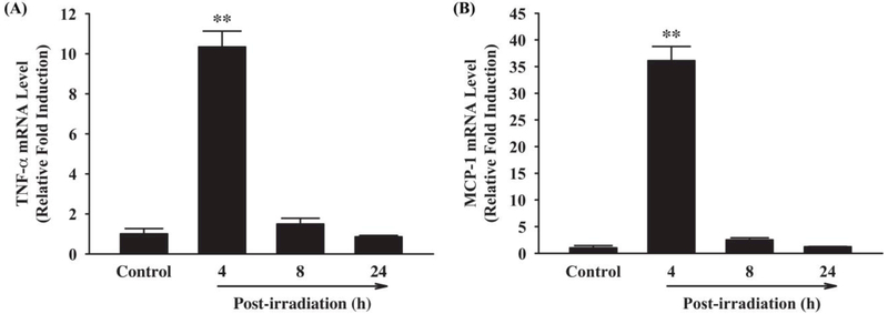
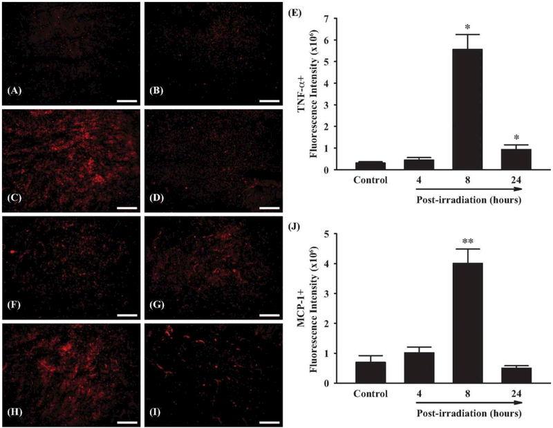
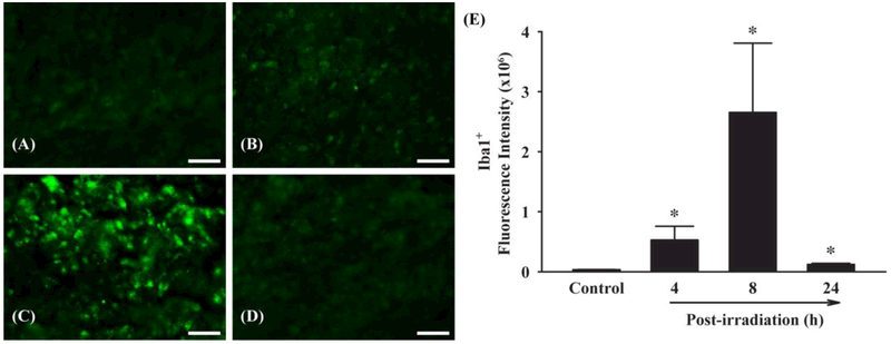
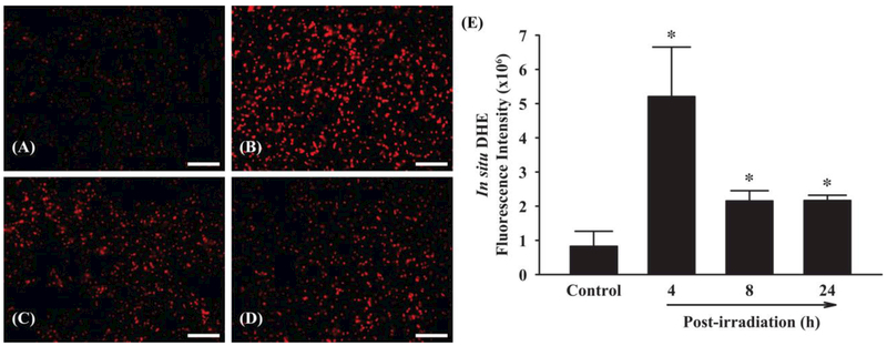
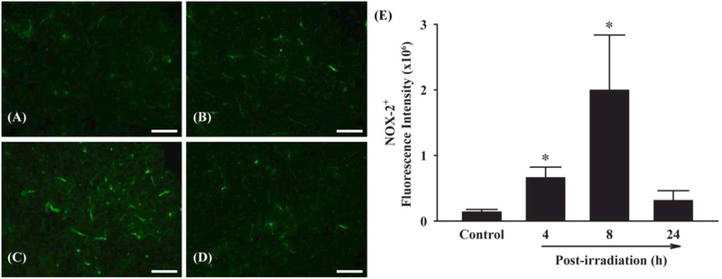
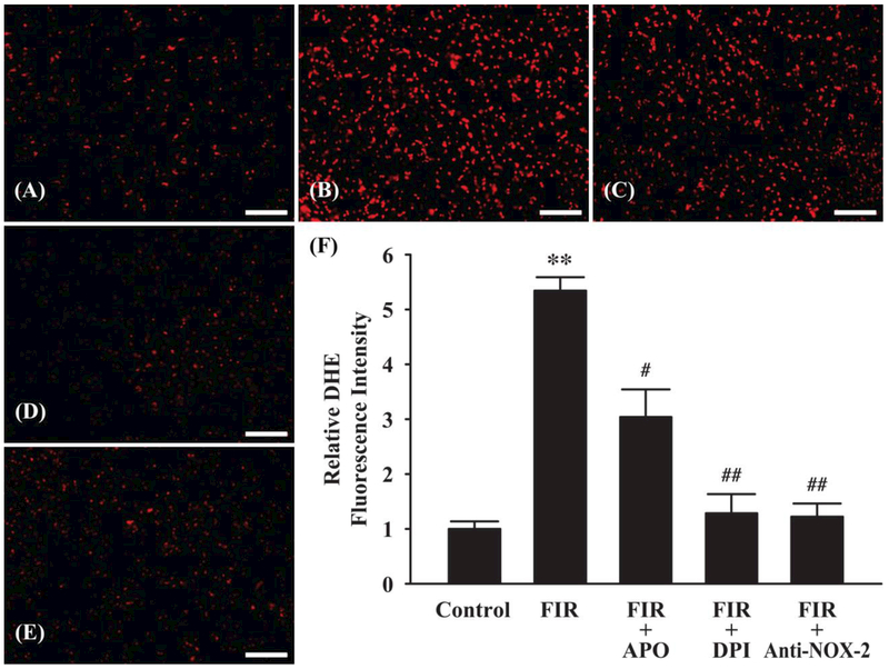
Materials and methods: C57BL/6 mice received either fractionated whole brain irradiation or sham-irradiation. The mRNA expression levels of pro-inflammatory mediators, such as TNF-α and MCP-1, were determined by quantitative real-time RT-PCR. The protein expression levels of TNF-α, MCP-1, NOX-2 and Iba1 were detected by immunofluorescence staining. The levels of ROS were visualized by in situ DHE fluorescence staining.
Results: A significant up-regulation of mRNA and protein expression levels of TNF-α and MCP-1 was observed in irradiated mouse brains. Additionally, immunofluorescence staining of Iba1 showed a marked increase of microglial activation in mouse brain after irradiation. Moreover, in situ DHE fluorescence staining revealed that fractionated whole brain irradiation significantly increased production of ROS. Furthermore, a significant increase in immunoreactivity of NOX-2 was detected in mouse brain after irradiation. On the contrary, an enhanced ROS generation in mouse brain after irradiation was markedly attenuated in the presence of NOX inhibitors or NOX-2 neutralizing antibody.
Conclusions: These results suggest that NOX-2 may play a role in fractionated whole brain irradiation-induced pro-oxidative and pro-inflammatory pathways in mouse brain.
Keywords: Fractionated whole brain irradiation; NOX-2; ROS; inflammation.
Conflict of interest statement
Declaration of Interest
The authors report no conflicts of interest.
Figures
References
-
- Akiyama K, Tanaka R, Sato M, Takeda N. 2001. Cognitive dysfunction and histological findings in adult rats one year after whole brain irradiation. Neurol Med Chir. 41:590–598. - PubMed
-
- Ashpole NM, Warrington JP, Mitschelen MC, Yan H, Sosnowska D, Gautam T, Farley JA, Csiszar A, Ungvari Z, Sonntag WE. Systemic influences contribute to prolonged microvascular rarefaction after brain irradiation: a role for endothelial progenitor cells. Am J Physiol Heart Circ Physiol. 2014;307(6):H858–868. - PMC - PubMed
-
- Baluchamy S, Zhang Y, Ravichandran P, Ramesh V, Sodipe A, Hall JC, Jejelowo O, Gridley DS, Wu H, Ramesh GT. 2010. Differential oxidative stress gene expression profile in mouse brain after proton exposure. In Vitro Cell Dev Biol Anim. 46:718–725. - PubMed
-
- Baluna RG, Eng TY, Thomas CR. 2006. Adhesion molecules in radiotherapy. Radiat Res. 166:819–831. - PubMed
-
- Barlind A, Karlsson N, Björk-Eriksson T, Isgaard J, Blomgren K. 2010. Decreased cytogenesis in the granule cell layer of the hippocampus and impaired place learning after irradiation of the young mouse brain evaluated using the IntelliCage platform. Exp Brain Res. 201:781–787. - PubMed
MeSH terms
Substances
Grants and funding
LinkOut - more resources
Full Text Sources
Other Literature Sources
Miscellaneous