. 2017 Sep 7;13(9):e1006549.
doi: 10.1371/journal.ppat.1006549. eCollection 2017 Sep.

Staphylococcal enterotoxin-like X (SElX) is a unique superantigen with functional features of two major families of staphylococcal virulence factors

Affiliations

Staphylococcal enterotoxin-like X (SElX) is a unique superantigen with functional features of two major families of staphylococcal virulence factors

Ries J Langley et al. PLoS Pathog. .

Abstract

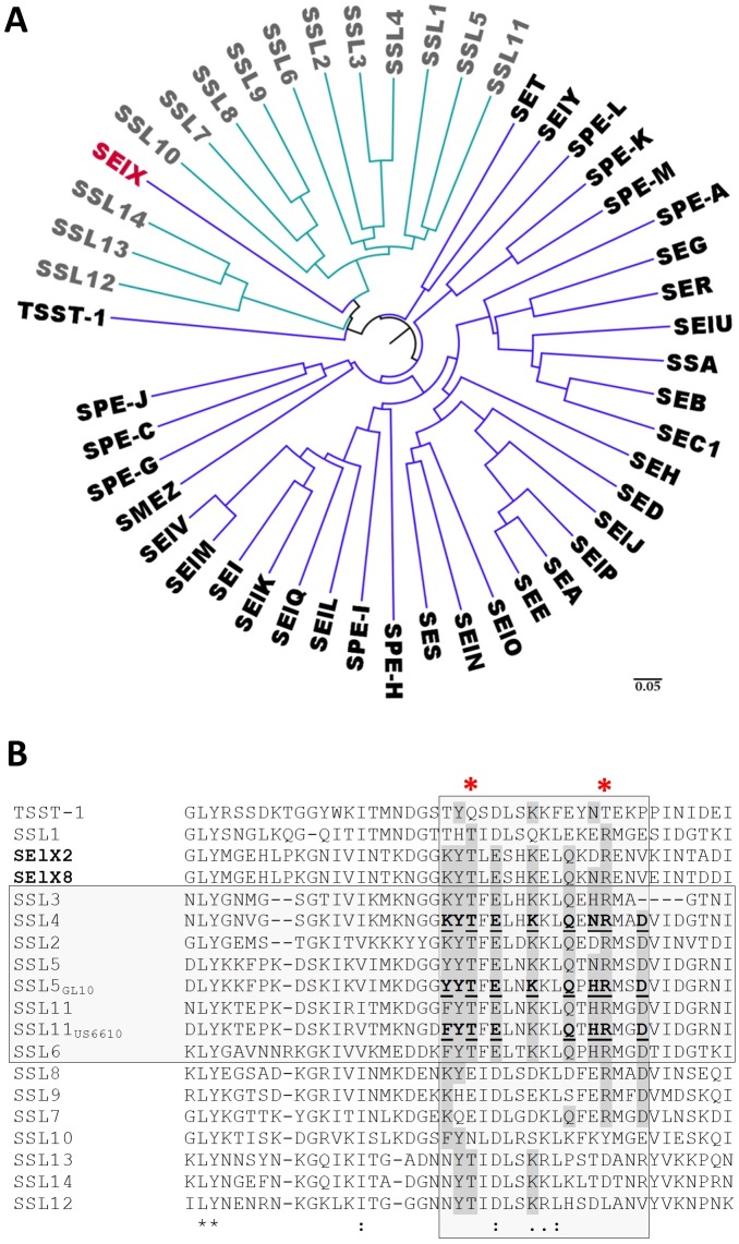
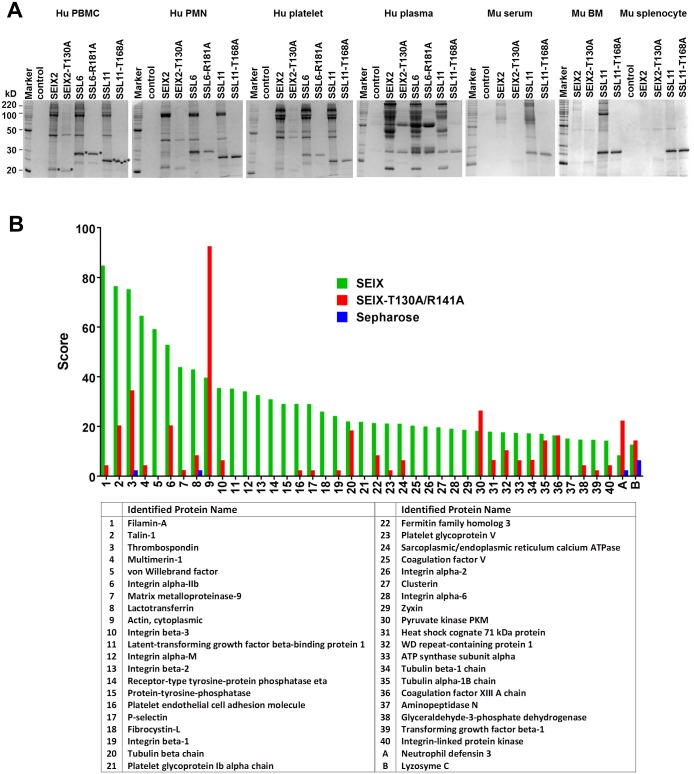
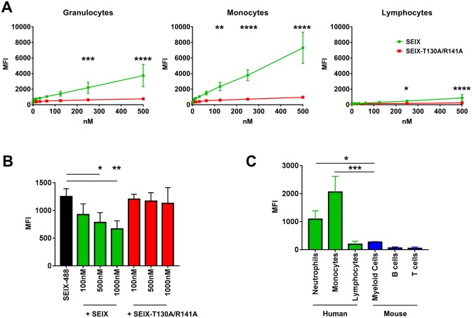
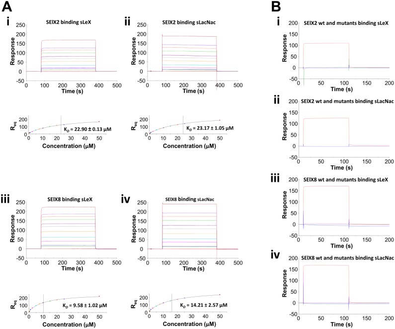
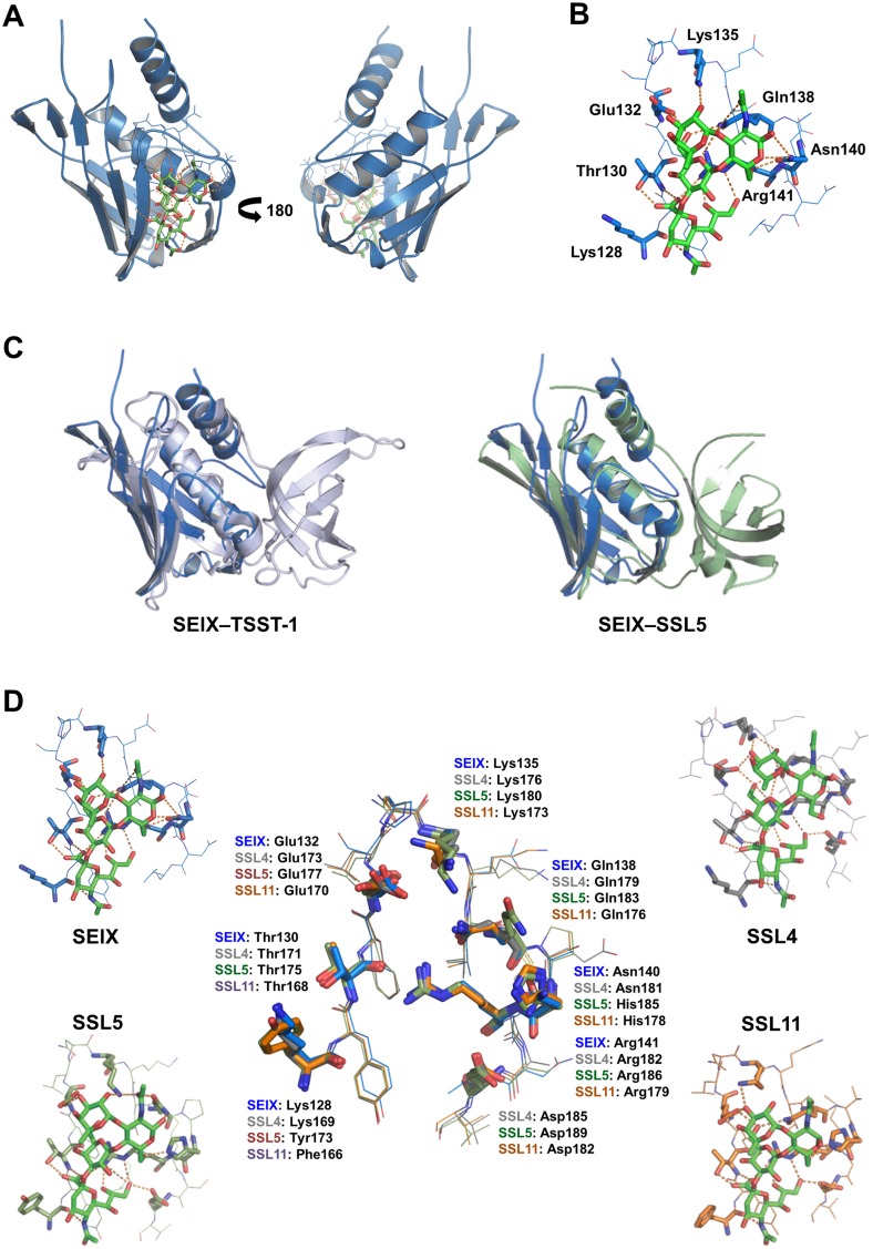
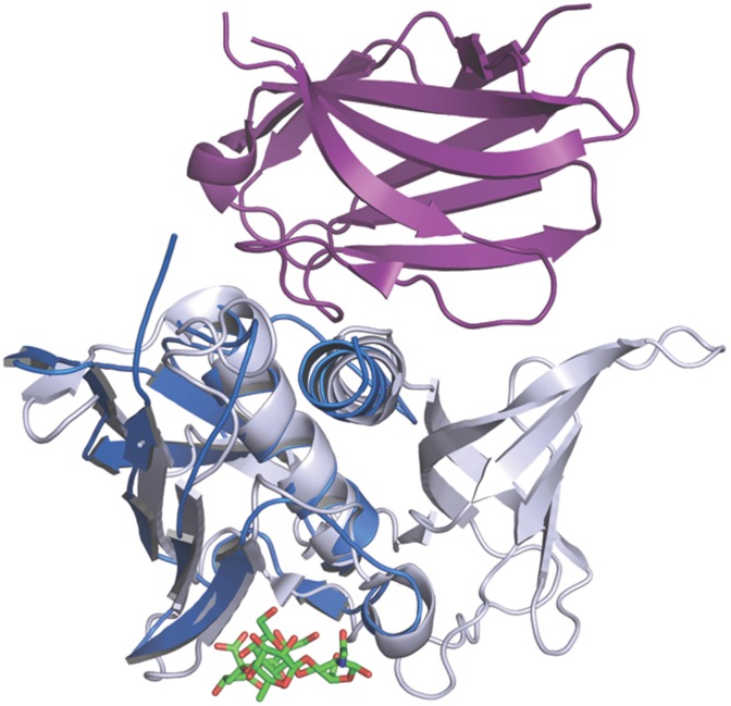
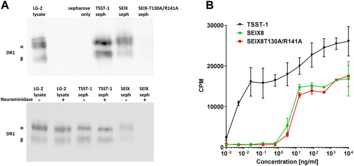
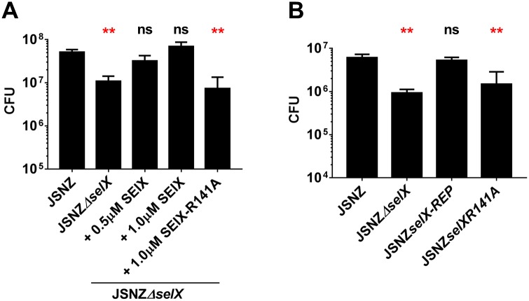
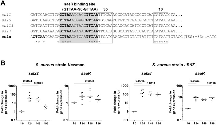
Staphylococcus aureus is an opportunistic pathogen that produces many virulence factors. Two major families of which are the staphylococcal superantigens (SAgs) and the Staphylococcal Superantigen-Like (SSL) exoproteins. The former are immunomodulatory toxins that induce a Vβ-specific activation of T cells, while the latter are immune evasion molecules that interfere with a wide range of innate immune defences. The superantigenic properties of Staphylococcal enterotoxin-like X (SElX) have recently been established. We now reveal that SElX also possesses functional characteristics of the SSLs. A region of SElX displays high homology to the sialyl-lactosamine (sLacNac)-specific binding site present in a sub-family of SSLs. By analysing the interaction of SElX with sLacNac-containing glycans we show that SElX has an equivalent specificity and host cell binding range to the SSLs. Mutation of key amino acids in this conserved region affects the ability of SElX to bind to cells of myeloid origin and significantly reduces its ability to protect S. aureus from destruction in a whole blood killing (WBK) assay. Like the SSLs, SElX is up-regulated early during infection and is under the control of the S. aureus exotoxin expression (Sae) two component gene regulatory system. Additionally, the structure of SElX in complex with the sLacNac-containing tetrasaccharide sialyl Lewis X (sLeX) reveals that SElX is a unique single-domain SAg. In summary, SElX is an 'SSL-like' SAg.


Conflict of interest statement

The authors declare that there are no financial, personal, or professional interests that could be construed to have influenced this work.

Figures

Fig 1
Fig 1. Comparison of SElX with SAgs and SSLs.
(A) Phylogenetic analysis of the SAgs and SSLs of S. aureus. The Phylogenetic tree created using FigTree (v1.4.2) from an amino acid alignment of the staphylococcal SAgs and SSLs generated using Clustal Omega (EMBL-EBI). SAgs are shown in black text and the SSLs in grey text. SElX is shown in red. (B) Amino acid sequence alignment of the two SElX variants used in this study, SElX2 and SElX8 (in bold), with the SSLs in the region of the sialylated glycan-dependent binding site. The glycan binding SSL subfamily is highlighted by the horizontal grey box and the region of the 17 amino acid glycan binding site is highlighted by the vertical grey box. Residues that have been experimentally determined to interact with the sialylated glycan are shown in bold type with those that hydrogen bond to the glycan underlined. Residues with homology to these amino acids are highlighted in dark grey. The conserved Threonine (T) and Arginine (R) residues mutated to affect sialylated glycan binding are indicated by the red asterisks.
Fig 2
Fig 2. Analysis of host binding by affinity precipitation.
(A) SDS-PAGE (12.5%) run under reducing and denaturing conditions of proteins from the various indicated human and mouse sources isolated by affinity with SElX2, SElX2-T130A, SSL6, SSL6-R181A, SSL11, or SSL11-T168A coupled to sepharose. Sepharose alone (control) was used as a control for non-specific binding. * indicates SELX/SSL that has dissociated from the sepharose. Marker is BenchMark Protein Ladder (Life Technologies). (B) The 40 top scoring leukocyte proteins identified by SElX-sepharose affinity binding are shown by descending rank in the bar graph and listed in order in the accompanying table. The ranking is based on the Unused Score (taken from S1 Table) given to each uniquely identified protein as calculated by the mass spectrometry analysis software ProteinPilot 5.0 (AB Sciex Pte. Ltd). The Unused Score indicates how much of the Total Score is unique to the particular protein hit. The Total Score is the sum of the Contrib values (contrib = the highest scoring peptide match for a peptide sequence) and determines the overall confidence for the protein identification. A and B are the Unused Scores of additional proteins identified in both the SElX-T130A/R141A and sepharose control samples with their corresponding Unused Scores from the SElX sample.
Fig 3
Fig 3. Analysis of host binding by flow cytometry.
(A) Median fluorescence intensity (MFI) of a two-fold dilution series of SElX-488 (green line) or SElX-T130A/R141A (red line) binding to human leukocytes with cell populations gated as granulocytes, monocytes, and lymphocytes based on size and granularity. Each data point represents the mean ± SD of three separate experiments using three individual donors. Comparison of the two proteins binding each cell population was performed in Graphpad Prism using two way analysis of variance (ANOVA) (p<0.0001) with Sidak’s multiple comparisons test: * p = 0.0336; ** p = 0.0037; ***p = 0.0005; **** p < 0.0001. (B) Binding of 100nM SElX-488 to human granulocytes (black bar) and in the presence of increasing concentrations of SElX (Green bars) or SElX-T130A/R141A (red bars). The MFI is the mean ± SD of three experiments performed using three separate human donors. The column data were compared by two-tailed paired t-tests: *p = 0.0333; ** p = 0.0038. (C) Comparison of the binding of 100nM SElX-488 to human and mouse leukocyte populations. The MFI is the mean ± SD of three experiments performed using three separate human donors or two experiments on n = 1 mouse per experiment and is the MFI (SElX-488 stained cells) minus MFI (unstained control population). Data compared by one way ANOVA (p<0.0001) with Tukey’s multiple comparisons test: * p = 0.0189; *** p = 0.0005.
Fig 4
Fig 4. Determination of SElX2 and SElX8 binding to sLeX and sLacNac by surface plasmon resonance.
(A) Quantitative measure of SElX2 and SElX8 binding to sLeX and sLacNac. Binding responses at equilibrium (Req) are shown against the concentration and fitted to a steady-state affinity binding model to calculate an equilibrium affinity constant (KD). (i) sLeX sensor chip binding and equilibrium binding analysis of 0.25 to 50 μM SElX2 in duplicate. (ii) sLacNac sensor chip binding and equilibrium binding analysis of 0.25 to 50 μM SElX2 in duplicate. (iii) sLeX sensor chip binding and equilibrium binding analysis of 0.25 to 50 μM SElX8 in duplicate. (iv) sLacNac sensor chip binding and equilibrium binding analysis of 0.25 to 50 μM SElX8 in duplicate. (B) Comparison of SElX and its glycan-binding mutants to sLeX and sLacNac. (i) sLeX sensor chip binding SElX2 (red), SElX2-T130A (pink), SElX2-R141A (green), and SElX2-T130A/R141A (blue) at 20 μM. (ii) sLacNac sensor chip binding SElX2 (red), SElX2-T130A (pink), SElX2-R141A (green), and SElX2-T130A/R141A (blue) at 20 μM. (iii) sLeX sensor chip binding SElX8 (red), SElX8-T130A (pink), SElX8-R141A (green), and SElX8-T130A/R141A (blue) at 20 μM. (iv) sLacNac sensor chip binding SElX8 (red), SElX8-T130A (pink), SElX8-R141A (green), and SElX8-T130A/R141A (blue) at 20 μM. The plots shown are representative of three independent experiments where each experiment was performed in duplicate. The affinity (KD) values are expressed as mean ± SD of the repeats.
Fig 5
Fig 5. The structural analysis of SElX.
(A) crystal structure of SElX8 (cyan) in complex with sLeX (green) shown from the left of the glycan binding site (left panel) and from the right of the glycan binding site (right panel). (B) The sialylated glycan-binding site of SElX8 (blue) showing the residues that hydrogen-bond (yellow dotted lines) with sLeX (green). The side chains that interact with sLeX are labelled and shown in bold. The components of sLeX are labelled as follows: N-Acetylneuraminic Acid (S); galactose (G); fucose (Fuc); and N-Acetylglucosamine (N). (C) Structural overlay of SElX8 (blue) with the SAg TSST-1 (silver) and SSL5 (sage). (D) Comparison of sialyl Lewis X (sLeX) (in green) bound in the glycan binding sites of SElX8 (blue), SSL4 (silver), SSL5 (sage), and SSL11 (orange). The side chains of residues that hydrogen-bond with sLeX are shown in bold. An overlay of these binding sites (centre) shows the conservation of residues that interact with sLeX.
Fig 6
Fig 6. Comparison of SElX with TSST-1 in complex with human Vβ2.
The structure of SElX (blue) with sLeX bound (in green) is overlaid with the structure of TSST-1 (silver) in complex with the Vβ2 region (purple) of a human TCR molecule (PDB: 2IJ0).
Fig 7
Fig 7. Association of the SElX glycan-binding site with MHC class II and T cell activation.
(A) Binding of SElX to MHC class II. Upper panel—immunoassay detecting for DR1 isolated from LG-2 cell lysate by TSST-1, SElX, or SElX-T130A/R141A coupled to sepharose. Lower panel—immunoassay detecting for DR1 isolated from LG-2 cells lysates treated ± with neuraminidase by TSST-1 or SElX coupled to sepharose. LG-2 lysate is run as a control to indicate the α and β chains of DR1. Sepharose only is a control for non-specific binding. (B) Effect of the SElX glycan-binding site on superantigen activity. Proliferation of human PBMCs by SElX, SElX-T130A/R141A, and TSST-1 measured by the incorporation as counts per minute (cpm) of 3H-thymindine. The data (mean ± SD) is a representative of the PBMC assay which was performed in triplicate on cells isolated from at least three healthy individuals.
Fig 8
Fig 8. Contribution of SElX to S. aureus survival in whole blood.
(A) Whole blood killing (WBK) of S. aureus strain JSNZ or JSNZΔselX +/- recombinant SElX (0.5 or 1.0 μM) or SElX-R141A at 1.0 μM. Bacterial survival was determined by CFU enumeration after 20 hr co-incubation. (B) WBK of S. aureus JSNZ, JSNZΔselX, JSNZselxR141A, or JSNZΔselxREP. Bacterial survival was determined by CFU enumeration after 20 hr co-incubation. The graphs are a representative of three independent experiments performed on three individual donors. Data is the mean ± SD of duplicate tests enumerated in triplicate. Statistics were performed using Graphpad Prism. Kruskal—Wallis one way ANOVA was performed (p = 0.0005) and comparisons between samples and JSNZ were made using Dunn's Multiple Comparison Test (** p< 0.01, ns = not significant).
Fig 9
Fig 9. Expression analysis of selX.
(A) Alignment of the region upstream from selX with the known upstream regulatory regions of ssl1, ssl7, ssl9, and ssl11. The -10 and -35 promoter elements are boxed and the region of homology to the saeR binding site (GTTAA-n6-GTTAA) is highlighted in grey. Conserved residues are indicated with an asterisk. (B). Relative selX transcript levels in S. aureus isolated from abscesses (n = 6) removed at 24 hr, 48 hr, or 96 hr after subcutaneous infection of mice with 5x10⁶ S. aureus Newman or JSNZ. selx, saeR expression normalized to reference genes gyrB and ftsZ and compared to in vitro expression of the inoculum. Each data point is the mean of triplicate values from an individual abscess, with the median of these mean values shown. Statistics were performed using Graphpad Prism. Kruskal—Wallis one way ANOVA was performed and comparisons between the in vivo samples and the inoculum were made using Dunn's Multiple Comparison Test. The p value of significantly different groups is shown.

Similar articles

Cited by

References

    1. Tong SC, Chen L, Fowler V Jr. Colonization, pathogenicity, host susceptibility, and therapeutics for Staphylococcus aureus: what is the clinical relevance? Seminars in Immunopathology. 2012;34(2):185–200. doi: 10.1007/s00281-011-0300-x - DOI - PMC - PubMed
    1. Williamson DA, Heffernan H. The changing landscape of antimicrobial resistance in New Zealand. N Z Med J. 2014;127(1403):41–54. - PubMed
    1. Wertheim HF, Melles DC, Vos MC, van Leeuwen W, van Belkum A, Verbrugh HA, et al. The role of nasal carriage in Staphylococcus aureus infections. Lancet Infect Dis. 2005;5(12):751–62. doi: 10.1016/S1473-3099(05)70295-4 - DOI - PubMed
    1. von Eiff C, Becker K, Machka K, Stammer H, Peters G. Nasal Carriage as a Source of Staphylococcus aureus Bacteremia. New England Journal of Medicine. 2001;344(1):11–6. doi: 10.1056/NEJM200101043440102 - DOI - PubMed
    1. Wertheim HF, Vos MC, Ott A, van Belkum A, Voss A, Kluytmans JA, et al. Risk and outcome of nosocomial Staphylococcus aureus bacteraemia in nasal carriers versus non-carriers. Lancet. 2004;364(9435):703–5. - PubMed