

Cerebrovascular gene expression in spontaneously hypertensive rats
- PMID: 28880918
- PMCID: PMC5589213
- DOI: 10.1371/journal.pone.0184233
Cerebrovascular gene expression in spontaneously hypertensive rats
Abstract
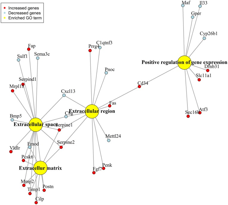
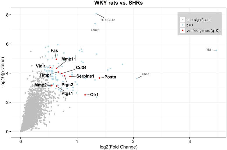
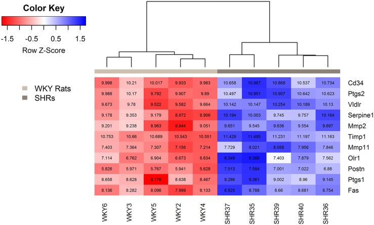
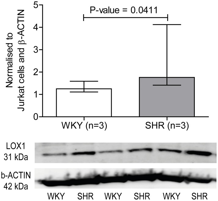
Hypertension is a hemodynamic disorder and one of the most important and well-established risk factors for vascular diseases such as stroke. Blood vessels exposed to chronic shear stress develop structural changes and remodeling of the vascular wall through many complex mechanisms. However, the molecular mechanisms involved are not fully understood. Hypertension-susceptible genes may provide a novel insight into potential molecular mechanisms of hypertension and secondary complications associated with hypertension. The aim of this exploratory study was to identify gene expression differences in the middle cerebral arteries between 12-week-old male spontaneously hypertensive rats and their normotensive Wistar-Kyoto rats using an Affymetrix whole-transcriptome expression profiling. Quantitative PCR and western blotting were used to verify genes of interest. 169 genes were differentially expressed in the middle cerebral arteries from hypertensive compared to normotensive rats. The gene expression of 72 genes was decreased and the gene expression of 97 genes was increased. The following genes with a fold difference ≥1.40 were verified by quantitative PCR; Postn, Olr1, Fas, Vldlr, Mmp2, Timp1, Serpine1, Mmp11, Cd34, Ptgs1 and Ptgs2. The gene expression of Postn, Olr1, Fas, Vldlr, Mmp2, Timp1 and Serpine1 and the protein expression of LOX1 (also known as OLR1) were significantly increased in the middle cerebral arteries from spontaneously hypertensive rats compared to Wistar-Kyoto rats. In conclusion, the identified genes in the middle cerebral arteries from spontaneously hypertensive rats could be possible mediators of the vascular changes and secondary complications associated with hypertension. This study supports the selection of key genes to investigate in the future research of hypertension-induced end-organ damage.
Conflict of interest statement
Figures
References
-
- Iadecola C, Davisson RL. Hypertension and Cerebrovascular Dysfunction. Cell Metab. 2008;7: 476–484. doi: 10.1016/j.cmet.2008.03.010 - DOI - PMC - PubMed
-
- Chobanian A V., Bakris GL, Black HR, Cushman WC, Green LA, Izzo JL, et al. Seventh report of the Joint National Committee on Prevention, Detection, Evaluation, and Treatment of High Blood Pressure. Hypertension. 2003;42: 1206–1252. doi: 10.1161/01.HYP.0000107251.49515.c2 - DOI - PubMed
-
- Mozaffarian D, Benjamin EJ, Go AS, Arnett DK, Blaha MJ, Cushman M, et al. Heart Disease and Stroke Statistics-2016 Update: A Report From the American Heart Association. Circulation. 2016;133: e38–60. doi: 10.1161/CIR.0000000000000350 - DOI - PubMed
-
- Intengan HD, Schiffrin EL. Vascular remodeling in hypertension: roles of apoptosis, inflammation, and fibrosis. Hypertension. 2001;38: 581–7. doi: 10.1161/hy09t1.096249 - DOI - PubMed
-
- Renna NF, de Las Heras N, Miatello RM. Pathophysiology of vascular remodeling in hypertension. Int J Hypertens. 2013/08/24. 2013;2013: 808353 doi: 10.1155/2013/808353 - DOI - PMC - PubMed
MeSH terms
LinkOut - more resources
Full Text Sources
Other Literature Sources
Medical
Research Materials
Miscellaneous
