. 2017 Sep 7;12(9):e0184575.
doi: 10.1371/journal.pone.0184575. eCollection 2017.

ARF6 mediates nephrin tyrosine phosphorylation-induced podocyte cellular dynamics

Affiliations

ARF6 mediates nephrin tyrosine phosphorylation-induced podocyte cellular dynamics

Jamie S Lin et al. PLoS One. .

Abstract

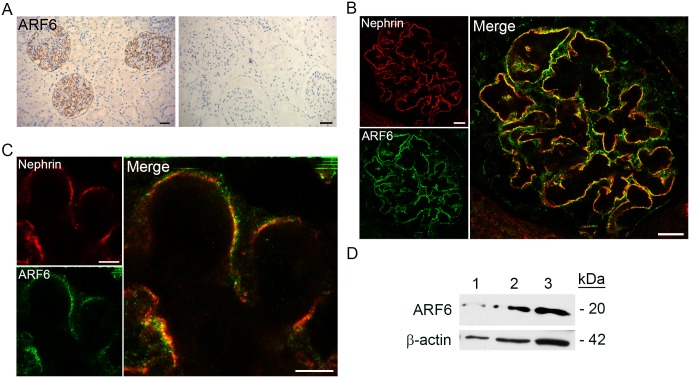
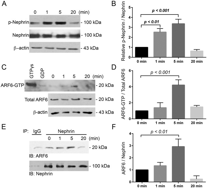
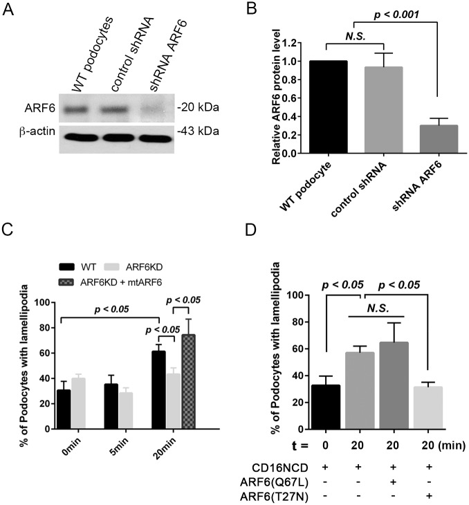
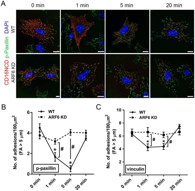
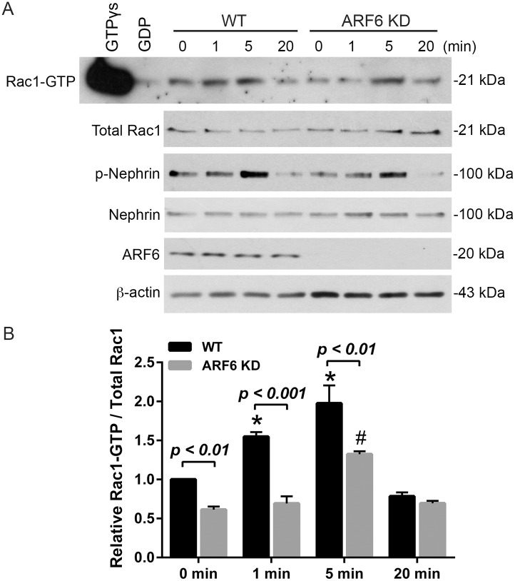
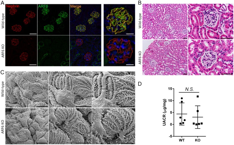
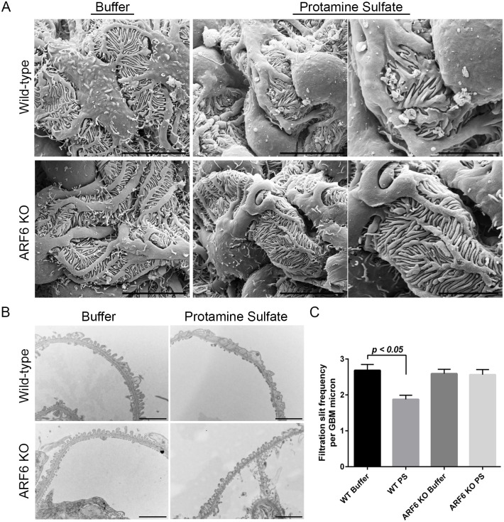
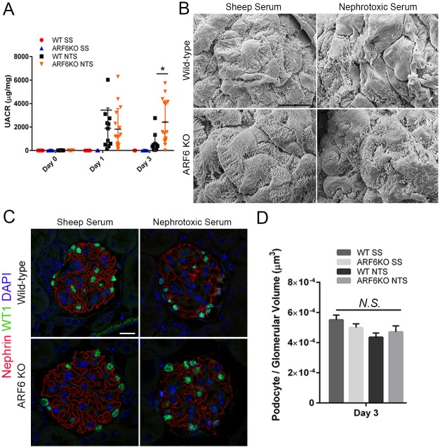
ADP-ribosylation factor 6 (ARF6) is a small GTPase necessary for regulating cellular structure, motility, and vesicle trafficking. In several cellular systems, ARF6 was shown to regulate actin dynamics in coordination with Rac1, a Rho small GTPase. We examined the function of ARF6 in the kidney podocyte because Rac1 was implicated in kidney diseases involving this cell. We found that ARF6 expression was enriched in human podocytes and that it modulated podocyte cytoskeletal dynamics through a functional interaction with nephrin, an intercellular junction protein necessary for podocyte injury-induced signaling requiring activation by tyrosine phosphorylation of its cytoplasmic domain. ARF6 was necessary for nephrin activation-induced ruffling and focal adhesion turnover, possibly by altering Rac1 activity. In podocyte-specific Arf6 (ARF6_PodKO) knockout mice, ARF6 deficiency did not result in a spontaneous kidney developmental phenotype or proteinuria after aging. However, ARF6_PodKO mice exhibited distinct phenotypes in two in vivo glomerular injury models. In the protamine sulfate perfusion model, which induced acute podocyte effacement, ARF6_PodKO mice were protected from podocyte effacement. In the nephrotoxic serum nephritis model, which induced immune-complex mediated injury, ARF6_PodKO mice exhibited aggravated proteinuria. Together, these observations suggest that while ARF6 is necessary for nephrin tyrosine phosphorylation-induced cytoskeletal dynamics in cultured podocytes, ARF6 has pleotropic podocyte roles in vivo, where glomerular injury-specific mechanisms might activate distinct signaling pathways that dictate whether ARF6 activity is beneficial or deleterious for maintaining the integrity of the glomerular filtration barrier.


Conflict of interest statement

Competing Interests: The authors have declared that no competing interests exist.

Figures

Fig 1
Fig 1. ARF6 is enriched in human glomerular podocytes.
A. Sections of normal human kidney obtained by nephrectomy were stained by immunohistochemistry for ARF6 (brown). Negative control (right). Scale bar 30 μm. B. Indirect immunofluorescence confocal microscopy was used to image normal human kidney tissue stained for ARF6 (green) and nephrin (red). Magnification x63. Scale bar 11μm. C. Stimulated emission depletion micrograph of normal human kidney tissue stained for ARF6 (green) and nephrin (red). Scale bar 5 μm. D. Immunoblot analysis of ARF6 from lysate prepared from 1) normal wild-type (WT) mouse kidney, 2) isolated rat glomeruli, and 3) human podocyte cell line. β-actin was used as loading control.
Fig 2
Fig 2. Nephrin tyrosine phosphorylation increased ARF6 activity and nephrin-ARF6 association.
A. Immunoblot (IB) of total nephrin and tyrosine-phosphorylated nephrin (p-Nephrin) using the CD16/7-nephrin activation model in CD16/7-nephrin expressing human WT podocytes. B. Densitometry analysis of p-nephrin to total nephrin normalized to 0 minutes. Data are shown as mean ± SEM. C. Following nephrin induction in WT podocytes ARF6 activity was assessed by an ARF6 activation assay. GTPγs- and GDP-treated samples were used as positive and negative controls, respectively. D. Densitometry analysis of ARF6-GTP to total ARF6 normalized to 0 minutes. E. CD16/7-nephrin-ARF6 association was assessed by co-immunoprecipitation (IP) where nephrin was immunoprecipitated from podocyte lysates, and nephrin and ARF6 were detected by IB. IP using rabbit IgG was employed as control. F. Densitometry analysis of ARF6 normalized to nephrin. Assays were repeated in triplicate.
Fig 3
Fig 3. ARF6 is necessary for nephrin ligation-induced ruffling activity.
A. IB of ARF6 protein expression in human WT podocytes, WT podocytes transduced with shRNA control, or ARF6 shRNA. B. Densitometry analysis of ARF6 protein abundance normalized to β-actin. C. Percent of podocytes with positive anti-lamellipodin staining following activation of CD16/7-nephrin expressing WT and ARF6 depleted cells. Expression of mutant ARF6 (mtARF6, in which four same-sense mutations were placed within the wild type human ARF6 shRNA target region) was used to rescue ruffling activity in cultured activated CD16/7-nephrin expressing stable ARF6 KD podocytes. D. Percent of podocytes with positive anti-lamellipodin staining 20 minutes following activation of CD16/7 expressing WT, constitutively-active ARF6, ARF6(Q67L), or dominant-negative, ARF6(T27N) podocytes. Approximately 100 cells were evaluated per condition.
Fig 4
Fig 4. ARF6KD inhibits nephrin activation-induced dynamic focal adhesion turnover.
A. CD16/7-nephrin (red) expressing WT and stable ARF6-depleted (ARF6KD) human podocytes were used to assess focal adhesion (FA) turnover following CD16/7-nephrin induction. Indirect immunofluorescence microscopy of WT and ARF6KD cells showing CD16/7-nephrin (red) and FA, phosphorylated-paxillin (p-paxillin, green), at indicated time-points following nephrin activation. Magnification 63x. Scale bar 20 μm. B. Quantification of the number of mature FA (≥5 μm Feret’s Diameter) per 100 μm2 cell surface area identified by p-paxillin staining at indicated times following nephrin activation. C. Quantification of the number of mature FA (≥5 μm Feret’s Diameter) per 100 μm2 cell surface area identified by vinculin staining at indicated times following nephrin activation.
Fig 5
Fig 5. ARF6KD attenuated nephrin tyrosine phosphorylation-induced Rac1 activity.
A. Following nephrin activation in CD16/7-nephrin expressing human WT or ARF6KD podocytes, Rac1 activity was determined as described in Methods. GTPγs and GDP treated samples were used as positive and negative control, respectively. B. Densitometry analysis of Rac1-GTP to total Rac1 normalized to time 0 minutes.
Fig 6
Fig 6. Podocyte-specific ARF6 null mice have no obvious renal abnormalities.
The Arf6 locus was targeted by homologous recombination with loxP sites flanking exon 1 and 2. Floxed Arf6 mice were bred with NPHS2-Cre (podocin-Cre) mice to obtain podocyte-specific ARF6 knockout (Arf6f/f;Nphs2-CreTg/+) mice. Mice homozygous for floxed Arf6 allele, but lacking Nphs2-Cre allele were used as control (Arf6f/f;Nphs2-Cre+/+). A. Indirect immunofluorescence microscopy of nephrin (red) and ARF6 (green) staining demonstrated podocyte-specific ARF6 deletion. Magnification (left) x40, scale bar 60 μm; (right) x63, scale bar 20 μm. B. Mouse kidney tissue sections were stained with Periodic acid-Schiff and examined by light microscopy. Magnification (left) x20, scale bar 60 μm. Magnification (right) x60, scale bar 60 μm. C. Glomerular ultrastructure was assessed by scanning electron microscope. Magnification (left) x3000, scale bar 10 μm; (middle) x6000, scale bar 10 μm; (right) x10000, scale bar 5 μm. D. Urine albumin to creatinine ratio was determined for 12-month-old podocyte-specific ARF6KO and control mice.
Fig 7
Fig 7. Podocyte-specific ARF6 null mice were protected from protamine sulfate-induced foot process effacement.
A. Protamine sulfate (PS) perfusion was performed in WT (Arf6f/f; Nphs2-Cre+/+) and podocyte-specific ARF6 null (Arf6f/f;Nphs2-CreTg/+) mice. Hank’s balanced salt solution was used as control buffer perfusion. Podocyte morphology was evaluated by scanning electron microscope. Magnification (left) x6000, scale bar 10 μm; (middle) x6000, scale bar 10 μm; (right) x10000, scale bar 5 μm. B. Foot process alterations were evaluated by transmission electron microscope. Magnification x10000, scale bar 2 μm. C. The number of intercellular junctions per glomerular basement membrane (GBM) length was compared between all groups of mice (mean ± SEM). Results are representative of 4–6 mice per group.
Fig 8
Fig 8. Podocyte-specific ARF6 null mice have aggravated proteinuria following nephrotoxic serum nephritis glomerular injury.
A. Urine albumin to creatinine ratio (UACR) was determined for sheep serum or nephrotoxic serum (NTS) treated WT and podocyte-specific ARF6 null mice (mean ± SEM). Results are representative of 10–15 mice per group. B. Day 3 glomerular ultrastructure was assessed by scanning electron microscope. Magnification x3000. Scale bar 5 μm. C. Indirect immunofluorescence of mouse kidney tissue sections stained with anti-WT1 (green) and anti-nephrin (red). Magnification x63. Scale bar 20 μm. D. Podocyte number per glomerular volume (μm3) in all treatment groups.

References

    1. Mundel P, Shankland SJ. Podocyte biology and response to injury. Journal of the American Society of Nephrology: JASN. 2002;13(12):3005–15. - PubMed
    1. Oh J, Reiser J, Mundel P. Dynamic (re)organization of the podocyte actin cytoskeleton in the nephrotic syndrome. Pediatric nephrology (Berlin, Germany). 2004;19(2):130–7. - PubMed
    1. Ichikawa I, Ma J, Motojima M, Matsusaka T. Podocyte damage damages podocytes: autonomous vicious cycle that drives local spread of glomerular sclerosis. Current opinion in nephrology and hypertension. 2005;14(3):205–10. - PubMed
    1. Wharram BL, Goyal M, Wiggins JE, Sanden SK, Hussain S, Filipiak WE, et al. Podocyte depletion causes glomerulosclerosis: diphtheria toxin-induced podocyte depletion in rats expressing human diphtheria toxin receptor transgene. Journal of the American Society of Nephrology: JASN. 2005;16(10):2941–52. doi: 10.1681/ASN.2005010055 - DOI - PubMed
    1. Patrakka J, Tryggvason K. New insights into the role of podocytes in proteinuria. Nature reviews Nephrology. 2009;5(8):463–8. doi: 10.1038/nrneph.2009.108 - DOI - PubMed