. 2017 Jun 13;8(31):50403-50414.
doi: 10.18632/oncotarget.18468. eCollection 2017 Aug 1.

Phospho-AXL is widely expressed in glioblastoma and associated with significant shorter overall survival

Affiliations

Phospho-AXL is widely expressed in glioblastoma and associated with significant shorter overall survival

Julia Onken et al. Oncotarget. .

Abstract

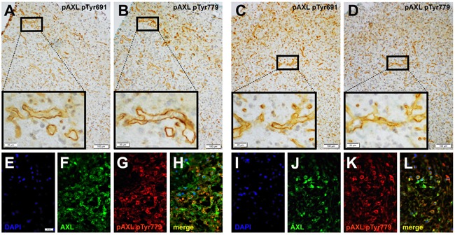
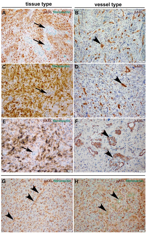
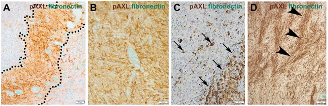
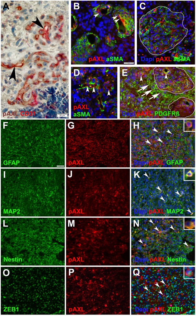
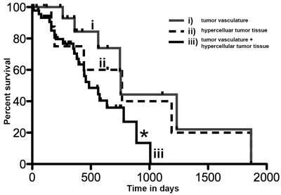
Receptor tyrosine kinase AXL (RTK-AXL) is regarded as a suitable target in glioblastoma (GBM) therapy. Since AXL kinase inhibitors are about to get approval for clinical use, patients with a potential benefit from therapy targeting AXL need to be identified. We therefore assessed the expression pattern of Phospho-AXL (P-AXL), the biologically active form of AXL, in 90 patients with newly diagnosed GBM, which was found to be detectable in 67 patients (corresponding to 74%). We identified three main P-AXL expression patterns: i) exclusively in the tumor vasculature (13%), ii) in areas of hypercellularity (35%), or iii) both, in the tumor vasculature and in hypercellular areas of the tumor tissue (52%). Pattern iii) is associated with significant decrease in overall survival (Hazard ratio 2.349, 95% confidence interval 1.069 to 5.162, *p=0.03). Our data suggest that P-AXL may serve as a therapeutic target in the majority of GBM patients.

Keywords: Pathology Section; glioblastoma multiforme (GBM); glomeroid tufts; overall survival; phospho-Axl (P-AXL); receptor tyrosine kinase AXL (AXL).


Conflict of interest statement

CONFLICTS OF INTEREST No conflicts of interest exists in the submission of the manuscript and the manuscript is approved by all authors for publication.

Figures

Figure 1
Figure 1
Immunohistochemical evaluation of serial sections of GBM tissue samples revealed the expression of both intracellular AXL phospho-sites - pTyr691 (A., C.) and pTyr779 (B., D.) - which were phosphorylated and expressed similarly in the tumor tissue. Immunofluorescent labeling of AXL (F., J.) and AXL phospho-site pTyr779 (G., K.) demonstrated colocalization (H., L.) of both antigens. Counterstaining was performed with 4’, 6-diamidino-2-phenylindoleI (DAPI; E., I.). (Scale bar: 100 μm A.-D., 20 μm A.-D.; inserts), 50 μm E.-L.)
Figure 2
Figure 2. Immunohistochemical double-staining of GBM tissue samples with antibodies against P-AXL (brown) and fibronectin (green) revealed three main P-AXL expression patterns
In samples classified as “tissue type”, P-AXL expression was mainly seen in areas of hypercellularity (A., C., E.). Tumor vessels which were highlighted by fibronectin staining were negative for P-AXL (arrows). Samples classified as “vessel type” showed exclusive P-AXL staining within the tumor vasculature (B., D., F.; arrowheads). The third pattern was characterized by P-AXL expression in both, the tumor tissue and vasculature (G., H.). P-AXL expression was detected in tubular (B., D., G.; arrowheads) and glomeruloid (F., H.; arrowheads) vascular proliferation. (Scale bar: 50 μm A.-D., F.-H.; 20 μm E.).
Figure 3
Figure 3
The expression pattern of P-AXL in hypercellular areas was further classified as focal (10-50%; A., C.) or diffuse (≥ 50%; B., D.). Focal expression was seen e.g. in pseudopalisades (A.; dashed line) or scattered clusters of tumor cells (C.; arrows). Other GBM samples demonstrated broad/global P-AXL expression (B., D.) which was pronounced in areas which often showed a typical herringbone-like pattern (D.; arrowheads). (Scale bar: 50 μm).
Figure 4
Figure 4
Immunohistochemical and immunofluorescent double-staining of GBM tissue samples revealed strong P-AXL expression in CD31 positive endothelial cells (A., B., D.; arrowheads), no colocalization with aSMA (B., C., D.), and partial colabeling with PDGFR-ß positive pericytes (E.; arrowheads) in microvascular proliferation. Glioma cells adjacent to microvascular proliferation (dashed lines) showed strong immunopositivity for PDGFR-ß (E.; arrows). P-AXL was further expressed by neoplastic glioma cells as noted by colabeling with GFAP (F.-H.; arrowheads), MAP2 (I.-K.; arrowheads), Nestin (L.-N.; arrowheads), and ZEB1 O.-Q.; arrowheads). (Scale bar: 20 μm A., 50 μm B.-Q.).
Figure 5
Figure 5. Kaplan Meier curve showing overall survival in days in GBM patients with P-AXL expression i) exclusively in the tumor vasculature, ii) in the hypercellular tumor tissue, or both iii) in tumor vasculature and hypercellular tumor tissue
The overall survival of patients with P-AXL expression in vital tumor tissue and tumor vessels (iii) was significantly reduced compared to patients with P-AXL expression in tumor tissue without vascular expression (ii). Log-rank (Mantel-Cox) Test *p = 0.0335, HR 2.349, 95% CI1.069 to 5.162.

References

    1. Ostrom QT, Gittleman H, Fulop J, Liu M, Blanda R, Kromer C, Wolinsky Y, Kruchko C, Barnholtz-Sloan JS. CBTRUS Statistical Report: Primary Brain and Central Nervous System Tumors Diagnosed in the United States in 2008-2012. Neuro Oncol. 2015;17(Suppl 4):iv1–iv62. doi: 10.1093/neuonc/nov189. - DOI - PMC - PubMed
    1. Reifenberger G, Wirsching HG, Knobbe-Thomsen CB, Weller M. Advances in the molecular genetics of gliomas - implications for classification and therapy. Nat Rev Clin Oncol. 2017;14:434–52. doi: 10.1038/nrclinonc.2016.204. - DOI - PubMed
    1. Stupp R, Dietrich PY, Ostermann Kraljevic S, Pica A, Maillard I, Maeder P, Meuli R, Janzer R, Pizzolato G, Miralbell R, Porchet F, Regli L, de Tribolet N, et al. Promising survival for patients with newly diagnosed glioblastoma multiforme treated with concomitant radiation plus temozolomide followed by adjuvant temozolomide. J Clin Oncol. 2002;20:1375–82. doi: 10.1200/JCO.2002.20.5.1375. - DOI - PubMed
    1. Stupp R, Mason WP, van den Bent MJ, Weller M, Fisher B, Taphoorn MJ, Belanger K, Brandes AA, Marosi C, Bogdahn U, Curschmann J, Janzer RC, Ludwin SK, et al. Radiotherapy plus concomitant and adjuvant temozolomide for glioblastoma. N Engl J Med. 2005;352:987–96. doi: 10.1056/NEJMoa043330. - DOI - PubMed
    1. Schwartzbaum JA, Fisher JL, Aldape KD, Wrensch M. Epidemiology and molecular pathology of glioma. Nat Clin Pract Neurol. 2006;2:494–503. doi: 10.1038/ncpneuro0289. - DOI - PubMed

LinkOut - more resources