. 2017 Mar 21;8(31):50582-50593.
doi: 10.18632/oncotarget.16410. eCollection 2017 Aug 1.

C/EBP-δ positively regulates MDSC expansion and endothelial VEGFR2 expression in tumor development

Affiliations

C/EBP-δ positively regulates MDSC expansion and endothelial VEGFR2 expression in tumor development

Yongfen Min et al. Oncotarget. .

Abstract

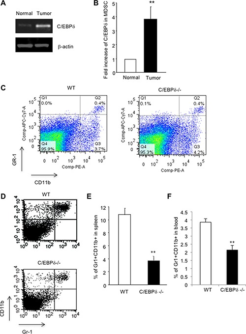
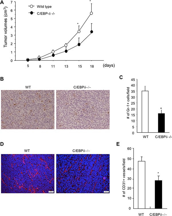
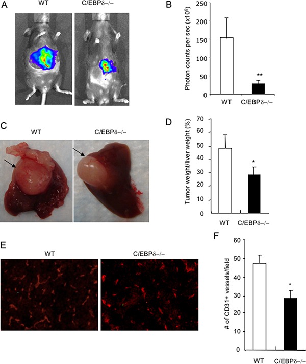
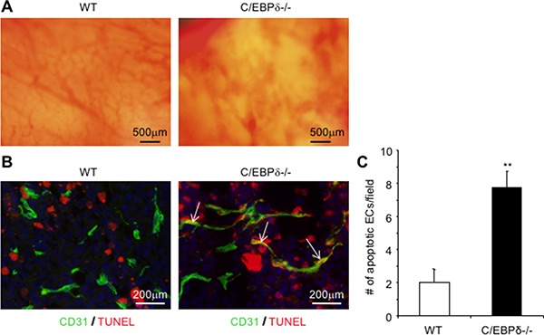
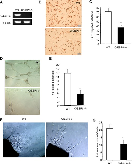
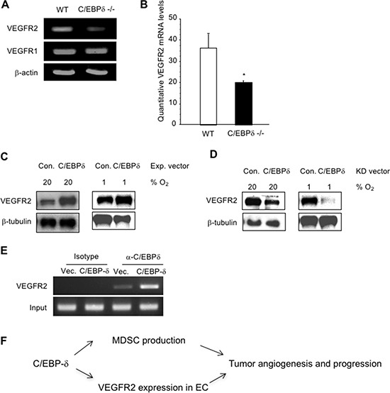
Vascular endothelial cells and Gr-1+CD11b+ myeloid derived suppressor cells (MDSCs) are two important components that constitute the tumor microenvironment. Targeting these cells offers the potential to halt tumor growth. In this study, we report a common mediator in C/EBP-δ that regulates both components and aids in tumor development. C/EBP-δ is elevated in tumor derived MDSCs. Interestingly, genetic deletion of C/EBP-δ in mice significantly impaired MDSC expansion in response to tumor progression, but it had no effect on Gr-1+CD11b+ cell production in normal development. It suggests a specific role of C/EBP-δ in emergency myelopoiesis under tumor conditions. Consistent with the pro tumor functions of MDSCs, loss of C/EBP-δ resulted in reduced tumor angiogenesis and tumor growth. Moreover, we found expression of C/EBP-δ in vascular endothelial cells. C/EBP-δ regulated cell motility, endothelial network formation and vascular sprouting. Notably, inactivation of C/EBP-δ in endothelial cells specifically inhibited the expression of VEGFR2 but not VEGFR1. Ectopic expression of C/EBP-δ increased and knockdown of the gene decreased VEGFR2 expression. C/EBP-δ is recruited to the promoter region of VEGFR2, indicative of transcriptional regulation. Collectively, this study has identified a positive mediator in C/EBP-δ, which regulates tumor induced MDSC expansion and VEGFR2 expression in endothelium. Considering the importance of MDSCs and endothelial cells in tumor progression, targeting C/EBP-δ may provide an interesting means for cancer therapy, killing two birds with one stone.

Keywords: C/EBP-δ; MDSCs; VEGFR2; angiogenesis; cancer.


Conflict of interest statement

CONFLICTS OF INTEREST The authors declare no conflicts of interest.

Figures

Figure 1
Figure 1. C/EBP-δ regulates tumor-induced expansion of MDSCs
Gr-1 and CD11b double positive cells were isolated by FACS sorting from spleens of C57Bl/6 mice with or without subcutaneous (s.c.) implantation of 3LL tumors. Total RNA was isolated and subjected to semi-quantitative RT-PCR (Panel A) and qPCR (Panel B) for C/EBP-δ expression. n = 3 mice per group, **p < 0.01. Gr-1CD11b double positive cells were analyzed by flow cytometry in spleens from age and sex matched C/EBP-δ null mice and littermate WT mice (Panel C) n = 5 mice per group. 5 × 10⁵ 3LL tumor cells were s.c. implanted into age and sex matched C/EBP-δ null mice and littermate WT mice for approximately 3 weeks. Single cell suspensions were made from spleens of mice with similar size tumors and analyzed by flow cytometry for Gr-1 and CD11b double positive cells (Panel D). The levels of Gr-1+CD11b+ cells in spleens (Panel E) and blood (Panel F) of tumor-bearing mice were plotted. n = 7 mice per group, **p < 0.01. Each experiment was repeated twice. Representative flow images are shown.
Figure 2
Figure 2. Genetic deletion of C/EBP-δ in mice results in a significant reduction of myeloid cell infiltration, tumor angiogenesis and tumor growth
5 × 10⁵ 3LL tumor cells were s.c. implanted into age and sex matched C/EBP-δ null mice and littermate WT mice. Tumor size was measured, calculated and plotted (Panel A). Tumor tissues were harvested from mice with similar size tumors and sectioned. Tissue sections were stained with a Gr-1 specific antibody and counterstained with hematoxylin. Brown color cells are Gr-1 positive cells (Panel B). Gr-1+ cells were counted in 10 randomly selected high power fields (Panel C). Tumor tissue sections were stained with a CD31 specific antibody (Panel D) and CD31+ vessels were counted in 10 randomly selected high power fields (Panel E). Data are expressed as mean ± SD. n = 10 mice per group, *p < 0.05.
Figure 3
Figure 3. Genetic deletion of C/EBP-δ in mice impairs tumor growth in a liver model
Small pieces of MC36-Luc tumor (1 mm3) were surgically implanted, one piece in each liver, in age and sex matched C/EBP-δ null mice and littermate WT mice. Tumors were imaged with bioluminescent imaging, and photon counts were used to measure tumor volume 3 weeks after tumor implantation (Panel A and B). Livers were harvested and grossly examined. Arrows pointed to the tumor (Panel C). Tumors were dissected and weighed. The percentage of tumor weight versus the weight of liver was plotted (Panel D). Tumor sections were stained with an anti CD31 antibody (Panel E), the number of CD31 positive vessels was counted in 10 randomly selected high power fields under microscopy (Panel F). Representative images are shown in each study. n = 7 mice per group and repeated once, **p < 0.01, *p < 0.05.
Figure 4
Figure 4. Loss of C/EBP-δ results in increased endothelial apoptosis and hemorrhagic vascular morphology in tumors
Dorsal skin vascular window chambers were established in WT and C/EBP-δ null mice, and 1 × 10⁵ 3LL tumor cells were injected into each window chamber. Tumor angiogenesis was imaged in live mice under microscopy 10 days after tumor cell implantation (Panel A). Tumor were harvested, sectioned and co-stained with an anti CD31 antibody (green) and TUNEL assay (red) (Panel B). The number of double positive cells was counted in 10 randomly selected high power fields under microscopy (Panel C). Representative images are shown. n = 5 mice per group. **p < 0.01.
Figure 5
Figure 5. C/EBP-δ is expressed in vascular endothelial cells, and regulates cell motility and angiogenesis
Pulmonary microvascular endothelial cells were isolated from age and sex matched C/EBP-δ null mice and littermate WT mice (pooled from 5 mice per group). Expression of C/EBP-δ in endothelial cells was examined by semi quantitative RT-PCR (Panel A). Endothelial cell migration was evaluated in a Transwell assay with seeding the primary murine endothelial cells in the upper chamber and addition of recombinant VEGF protein at 20 ng/ml in the bottom chamber (Panel B). The migrated cells were counted 5 hours later (Panel C). **p < 0.01. Murine endothelial cells were seeded on the top of Matrigel and incubated for 24 hours. Vascular network formation was imaged under microscopy (Panel D). The number of cross point of vascular structures was counted in 10 randomly selected fields under microscopy (Panel E). **p < 0.01. Aortas collected from age and sex matched WT and C/EBP-δ null mice (n = 3 mice per group) were cut into small pieces, embedded in fibrin gel and overlaid with EGM. The number of vascular sprouts was imaged and counted under microscopy after one week in culture (Panel F and G). **p < 0.01, *p < 0.05. All in vitro and ex vivo data were done in triplicates and collected from three independent experiments, and expressed as mean ± SD. Representative images are shown in each study.
Figure 6
Figure 6. C/EBP-δ binds to the promoter region of VEGFR2 and regulates its expression in endothelial cells
The mRNA levels of VEGFR1 in murine endothelial cells from WT and C/EBP-δ null mice were analyzed by semi quantitative RT-PCR (Panel A), and the mRNA levels of VEGFR2 were measured by both semi quantitative RT-PCR (Panel A) and qPCR (Panel B). *p < 0.05. HUVECs were transfected with either empty vector or C/EBP-δ expression vector for 24 hours. The cells were then cultured under either normoxia (20% O2) or hypoxia (1% O2) for another 24 hours. VEGFR2 protein levels were measured by Western blot (Panel C). HUVECs were transfected with either control shRNA or shRNA for C/EBP-δ for 24 hours. The cells were then cultured under either normoxia or hypoxia for another 24 hours. VEGFR2 protein levels were measured by Western blot (Panel D). HUVECs were transfected with either control vector or C/EBP-δ expression vector for 70 hours. ChIP assay was performed and the VEGFR2 promoter region was amplified by PCR (Panel E). Each experiment was repeated three times. Representative images are shown. Diagram of C/EBP-δ mediated tumor angiogenesis and MDSC (Panel F)expansion in tumor progression (Panel F).

References

    1. Balamurugan K, Wang JM, Tsai HH, Sharan S, Anver M, Leighty R, Sterneck E. The tumour suppressor C/EBPdelta inhibits FBXW7 expression and promotes mammary tumour metastasis. Embo J. 2010;29:4106–4117. - PMC - PubMed
    1. Balamurugan K, Sharan S, Klarmann KD, Zhang Y, Coppola V, Summers GH, Roger T, Morrison DK, Keller JR, Sterneck E. FBXW7alpha attenuates inflammatory signalling by downregulating C/EBPdelta and its target gene Tlr4. Nat Commun. 2013;4:1662. - PMC - PubMed
    1. Bjerregaard MD, Jurlander J, Klausen P, Borregaard N, Cowland JB. The in vivo profile of transcription factors during neutrophil differentiation in human bone marrow. Blood. 2003;101:4322–4332. - PubMed
    1. Chang W, Parra M, Centrella M, McCarthy TL. Interactions between CCAAT enhancer binding protein delta and estrogen receptor alpha control insulin-like growth factor I (igf1) and estrogen receptor-dependent gene expression in osteoblasts. Gene. 2005;345:225–235. - PubMed
    1. DeBusk LM, Boelte K, Min Y, Lin PC. Heterozygous deficiency of delta-catenin impairs pathological angiogenesis. J Exp Med. 2010;207:77–84. - PMC - PubMed