. 2017 Jun 8;8(32):53336-53351.
doi: 10.18632/oncotarget.18408. eCollection 2017 Aug 8.

Clinicopathological characteristics of ROS1- and RET- rearranged NSCLC in caucasian patients: Data from a cohort of 713 non-squamous NSCLC lacking KRAS/EGFR/HER2/BRAF/PIK3CA/ALK alterations

Affiliations

Clinicopathological characteristics of ROS1- and RET- rearranged NSCLC in caucasian patients: Data from a cohort of 713 non-squamous NSCLC lacking KRAS/EGFR/HER2/BRAF/PIK3CA/ALK alterations

Frédéric Dugay et al. Oncotarget. .

Abstract

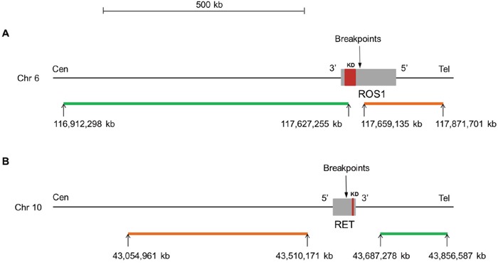
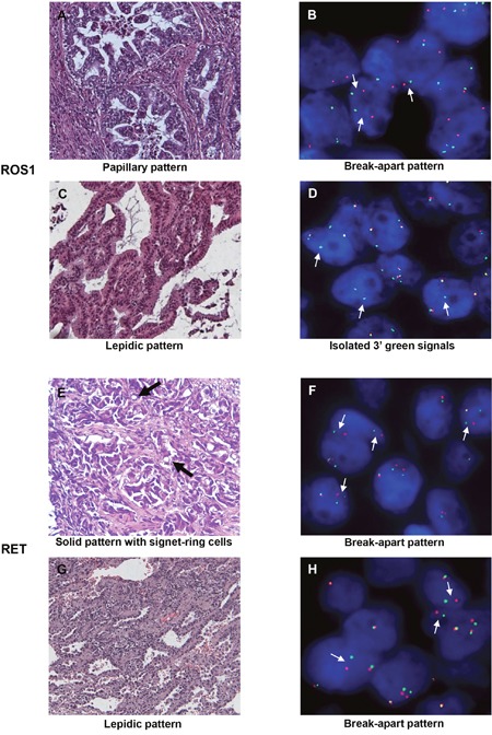
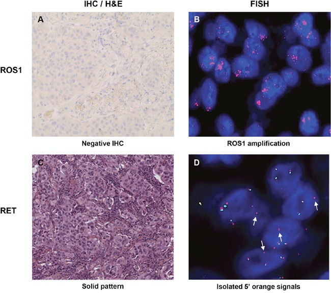
Targeted therapies have substantially changed the management of non-small cell lung cancer (NSCLC) patients with driver oncogenes. Given the high frequency, EGFR and ALK aberrations were the first to be detected and paved the way for tyrosine kinase inhibitor (TKI) treatments. Other kinases such as ROS1 and more recently RET have emerged as promising targets, and ROS1 and RET TKIs are already available for precision medicine. We screened a large cohort of 713 Caucasian non-squamous NSCLC patients lacking EGFR/KRAS/BRAF/HER2/PI3KCA/ALK aberrations for ROS1 and RET rearrangements using fluorescence in situ hybridization to determine the frequency and clinicopathological characteristics of ROS1- and RET-positive patients. Frequencies of ROS1 and RET rearrangements were 2.1% and 2.52%, respectively. Contrary to common belief, both ROS1 and RET rearrangements were detected in patients with a history of smoking, and the RET-positive patients were not younger than the negative patients. Moreover, RET but not ROS1 rearrangement was associated with the female gender. Nearly half of the ROS1-rearranged patients were successfully treated with ROS1 TKIs. In contrast, only 5/18 RET-positive patients received off-label RET TKIs. Two patients had stable disease, and three experienced disease progression. In addition to the 18 RET-positive cases, 10 showed isolated 5' signals. The clinical relevance is unknown but if the frequency is confirmed by other groups, the question whether these patients are eligible to TKIs will arise. More potent RET TKIs are under development and may improve the response rate in RET-positive patients. Therefore, we recommend the routine implementation of RET testing in non-squamous NSCLC patients, including those with a history of smoking.

Keywords: RET; ROS1; caucasian population; fusion genes; non-small cell lung cancer.


Conflict of interest statement

CONFLICTS OF INTEREST All authors have no conflicts of interest to disclose.

Figures

Figure 1
Figure 1
Map for ROS1 (A) and RET (B) dual color break-apart probes (ZytoLight® SPEC ROS1 and RET, ZytoVision). For both genes, the orange and green fluorochrome direct labeled probes hybridize proximal (5′ end) and distal (3′ end) to the genes, respectively. The known breakpoints in ROS1 are located in introns 31, 33 and 34 and are proximal to the kinase domain (exons 36 to 41). For RET, the known breakpoints are located in introns 10 and 11 and are proximal to the kinase domain (exons 12 to 19). The precise location of the genes on the chromosome, the size (kb) and orientation of the genes are indicated according to the Human assembly GRCh37/hg19. kb: kilobase pair; Cen: centromere; Tel: telomere; Chr: chromosome; KD: kinase domain.
Figure 2
Figure 2. Images of ROS1- or RET-rearranged lung adenocarcinoma
H&E staining (A, C, E, G) and corresponding FISH profiles (B, D, F, H) are shown. Thin white arrows indicate split signals or isolated 3′ green signals. Thick black arrows indicate signet-ring cells. H&E: hematoxylin and eosin (200x); FISH: fluorescence in situ hybridization.
Figure 3
Figure 3
Images of samples with ROS1 amplification (A-B) and atypical isolated 5′ signal pattern for RET (C-D). White arrows indicate isolated 5′ orange signals. IHC: immunohistochemistry; H&E: hematoxylin and eosin (200x); FISH: fluorescence in situ hybridization.

References

    1. Torre LA, Bray F, Siegel RL, Ferlay J, Lortet-Tieulent J, Jemal A. Global cancer statistics, 2012. CA Cancer J Clin. 2015;65:87–108. doi: 10.3322/caac.21262. - DOI - PubMed
    1. Saito M, Shiraishi K, Kunitoh H, Takenoshita S, Yokota J, Kohno T. Gene aberrations for precision medicine against lung adenocarcinoma. Cancer Sci. 2016;107:713–20. doi: 10.1111/cas.12941. - DOI - PMC - PubMed
    1. Soda M, Choi YL, Enomoto M, Takada S, Yamashita Y, Ishikawa S, Fujiwara S, Watanabe H, Kurashina K, Hatanaka H, Bando M, Ohno S, Ishikawa Y, et al. Identification of the transforming EML4-ALK fusion gene in non-small-cell lung cancer. Nature. 2007;448:561–6. doi: 10.1038/nature05945. - DOI - PubMed
    1. Shaw AT, Kim DW, Nakagawa K, Seto T, Crinó L, Ahn MJ, De Pas T, Besse B, Solomon BJ, Blackhall F, Wu YL, Thomas M, O’Byrne KJ, et al. Crizotinib versus chemotherapy in advanced ALK-positive lung cancer. N Engl J Med. 2013;368:2385–94. doi: 10.1056/NEJMoa1214886. - DOI - PubMed
    1. Rikova K, Guo A, Zeng Q, Possemato A, Yu J, Haack H, Nardone J, Lee K, Reeves C, Li Y, Hu Y, Tan Z, Stokes M, et al. Global survey of phosphotyrosine signaling identifies oncogenic kinases in lung cancer. Cell. 2007;131:1190–203. doi: 10.1016/j.cell.2007.11.025. - DOI - PubMed