

Role of mitochondrial dysfunction and dysregulation of Ca2+ homeostasis in the pathophysiology of insulin resistance and type 2 diabetes
- PMID: 28882140
- PMCID: PMC5588717
- DOI: 10.1186/s12929-017-0375-3
Role of mitochondrial dysfunction and dysregulation of Ca2+ homeostasis in the pathophysiology of insulin resistance and type 2 diabetes
Abstract
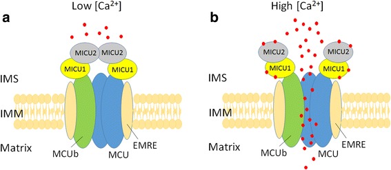
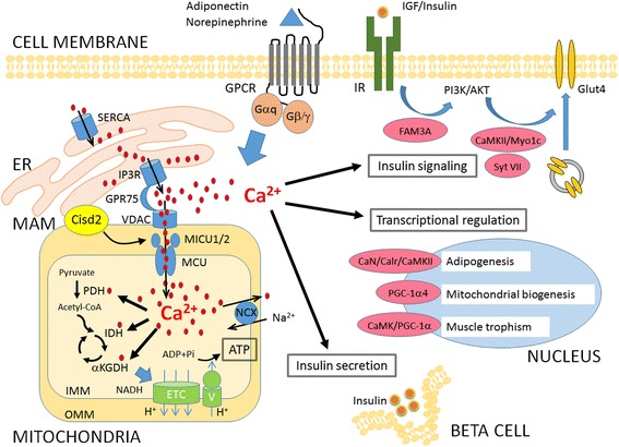
Metabolic diseases such as obesity, type 2 diabetes (T2D) and insulin resistance have attracted great attention from biomedical researchers and clinicians because of the astonishing increase in its prevalence. Decrease in the capacity of oxidative metabolism and mitochondrial dysfunction are a major contributor to the development of these metabolic disorders. Recent studies indicate that alteration of intracellular Ca2+ levels and downstream Ca2+-dependent signaling pathways appear to modulate gene transcription and the activities of many enzymes involved in cellular metabolism. Ca2+ uptake into mitochondria modulates a number of Ca2+-dependent proteins and enzymes participating in fatty acids metabolism, tricarboxylic acid cycle, oxidative phosphorylation and apoptosis in response to physiological and pathophysiological conditions. Mitochondrial calcium uniporter (MCU) complex has been identified as a major channel located on the inner membrane to regulate Ca2+ transport into mitochondria. Recent studies of MCU complex have increased our understanding of the modulation of mitochondrial function and retrograde signaling to the nucleus via regulation of the mitochondrial Ca2+ level. Mitochondria couple cellular metabolic state by regulating not only their own Ca2+ levels, but also influence the entire network of cellular Ca2+ signaling. The mitochondria-associated ER membranes (MAMs), which are specialized structures between ER and mitochondria, are responsible for efficient communication between these organelles. Defects in the function or structure of MAMs have been observed in affected tissue cells in metabolic disease or neurodegenerative disorders. We demonstrated that dysregulation of intracellular Ca2+ homeostasis due to mitochondrial dysfunction or defects in the function of MAMs are involved in the pathogenesis of insulin insensitivity and T2D. These observations suggest that mitochondrial dysfunction and disturbance of Ca2+ homeostasis warrant further studies to assist the development of therapeutics for prevention and medication of insulin resistance and T2D.
Keywords: Ca2+ homeostasis; Insulin resistance; Metabolic disease; Mitochondria-associated ER membranes; Mitochondrial calcium uniporter; Type 2 diabetes.
Conflict of interest statement
Ethics approval and consent to participate
Not applicable.
Consent for publication
Not applicable.
Competing interests
The authors declare that they have no competing interests.
Publisher’s Note
Springer Nature remains neutral with regard to jurisdictional claims in published maps and institutional affiliations.
Figures
Similar articles
-
Role of mitochondrial dysfunction and dysregulation of Ca(2+) homeostasis in insulin insensitivity of mammalian cells.Ann N Y Acad Sci. 2015 Sep;1350:66-76. doi: 10.1111/nyas.12838. Epub 2015 Jul 27. Ann N Y Acad Sci. 2015. PMID: 26214798 Review.
-
Mitochondrial VDAC, the Na+/Ca2+ Exchanger, and the Ca2+ Uniporter in Ca2+ Dynamics and Signaling.Adv Exp Med Biol. 2017;981:323-347. doi: 10.1007/978-3-319-55858-5_13. Adv Exp Med Biol. 2017. PMID: 29594867 Review.
-
Insight of the role of mitochondrial calcium homeostasis in hepatic insulin resistance.Mitochondrion. 2022 Jan;62:128-138. doi: 10.1016/j.mito.2021.11.007. Epub 2021 Nov 29. Mitochondrion. 2022. PMID: 34856389 Review.
-
Role of Endoplasmic Reticulum-Mitochondria Communication in Type 2 Diabetes.Adv Exp Med Biol. 2017;997:171-186. doi: 10.1007/978-981-10-4567-7_13. Adv Exp Med Biol. 2017. PMID: 28815530 Review.
-
Increased mitochondrial calcium uniporter in adipocytes underlies mitochondrial alterations associated with insulin resistance.Am J Physiol Endocrinol Metab. 2017 Dec 1;313(6):E641-E650. doi: 10.1152/ajpendo.00143.2016. Epub 2017 Aug 8. Am J Physiol Endocrinol Metab. 2017. PMID: 28790027 Free PMC article.
Cited by
-
Knowledge mapping of mitochondrial calcium uniporter from 2011 to 2022: A bibliometric analysis.Front Physiol. 2023 Jan 20;14:1107328. doi: 10.3389/fphys.2023.1107328. eCollection 2023. Front Physiol. 2023. PMID: 36744031 Free PMC article.
-
Therapeutic Strategies Targeting Mitochondrial Calcium Signaling: A New Hope for Neurological Diseases?Antioxidants (Basel). 2022 Jan 15;11(1):165. doi: 10.3390/antiox11010165. Antioxidants (Basel). 2022. PMID: 35052668 Free PMC article. Review.
-
Protein Kinases Signaling in Pancreatic Beta-cells Death and Type 2 Diabetes.Adv Exp Med Biol. 2021;1275:195-227. doi: 10.1007/978-3-030-49844-3_8. Adv Exp Med Biol. 2021. PMID: 33539017
-
Diabetes mellitus promotes susceptibility to periodontitis-novel insight into the molecular mechanisms.Front Endocrinol (Lausanne). 2023 Aug 16;14:1192625. doi: 10.3389/fendo.2023.1192625. eCollection 2023. Front Endocrinol (Lausanne). 2023. PMID: 37664859 Free PMC article. Review.
-
RAGE Differentially Altered in vitro Responses in Vascular Smooth Muscle Cells and Adventitial Fibroblasts in Diabetes-Induced Vascular Calcification.Front Physiol. 2021 Jun 7;12:676727. doi: 10.3389/fphys.2021.676727. eCollection 2021. Front Physiol. 2021. PMID: 34163373 Free PMC article.
References
-
- Yamaguchi T, Kanazawa I, Takaoka S, Sugimoto T. Serum calcium is positively correlated with fasting plasma glucose and insulin resistance, independent of parathyroid hormone, in male patients with type 2 diabetes mellitus. Metabolism. 2011;60(9):1334–1339. doi: 10.1016/j.metabol.2011.02.003. - DOI - PubMed
-
- Varadi A, Lebel L, Hashim Y, Mehta Z, Ashcroft SJ, Turner R. Sequence variants of the sarco (endo) plasmic reticulum Ca2+-transport ATPase 3 gene (SERCA3) in Caucasian type II diabetic patients (UK prospective diabetes study 48) Diabetologia. 1999;42(10):1240–1243. doi: 10.1007/s001250051298. - DOI - PubMed
Publication types
MeSH terms
Substances
LinkOut - more resources
Full Text Sources
Other Literature Sources
Medical
Miscellaneous
