. 2017 Sep 7;19(1):198.
doi: 10.1186/s13075-017-1407-9.

Estrogen attenuates the spondyloarthritis manifestations of the SKG arthritis model

Affiliations

Estrogen attenuates the spondyloarthritis manifestations of the SKG arthritis model

Hyemin Jeong et al. Arthritis Res Ther. .

Abstract

Background: Ankylosing spondylitis (AS) is a male-predominant disease, and radiographic evidence of damage is also more severe in males. Estrogen modulates immune-related processes such as T cell differentiation and cytokine production. This study aimed to evaluate the effect of estrogen on the disease activity of spondyloarthritis (SpA).

Methods: The effects of estrogen on the development of arthritis were evaluated by performing ovariectomy and 17β-estradiol (E2) pellet implantation in zymosan-treated SKG mice. Clinical arthritis scores were measured, and 18F-fluorodeoxyglucose (18F-FDG) small-animal positron emission tomography/computed tomography performed to quantify joint inflammation. The expression of inflammatory cytokines in joint tissue was measured.

Results: E2-treated mice showed remarkable suppression of arthritis clinically and little infiltration of inflammatory cells in the Achilles tendon and intervertebral disc. 18F-FDG uptake was significantly lower in E2-treated mice than in sham-operated (sham) and ovariectomized mice. Expression of TNF, interferon-γ, and IL-17A was significantly reduced in E2-treated mice, whereas expression of sclerostin and Dickkopf-1 was increased in E2-treated mice compared with sham and ovariectomized mice.

Conclusions: Estrogen suppressed arthritis development in SKG mice, a model of SpA. Results of this study suggest that estrogen has an anti-inflammatory effect on the spondyloarthritis manifestations of the SKG arthritis model.

Keywords: Estrogen; Mice; Spondyloarthritis.


Conflict of interest statement

Ethics approval

All animal studies were approved by the Institutional Animal Care and Use Committee at Samsung Medical Center.

Consent for publication

Not applicable.

Competing interests

The authors declare that they have no competing interests.

Publisher’s Note

Springer Nature remains neutral with regard to jurisdictional claims in published maps and institutional affiliations.

Figures
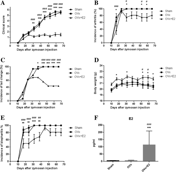
Fig. 1
Fig. 1
17β-estradiol (E2) treatment suppressed arthritis development in SKG mice. Clinical scores were markedly suppressed in E2-treated mice compared with mice in the sham-operated (Sham) and ovariectomized (OVx) groups after 31 days of zymosan injection. Clinical scores (a), the incidence of arthritis (b), the incidence of tail changes (c), body weight (d), and the incidence of blepharitis (e) in the three groups. Serum levels of E2 were measured by ELISA (f). Values are mean ± SEM (n = 16 mice per group); *P < 0.05, **P < 0.01, ***P < 0.001 (sham vs. OVx + E2); # P < 0.05; ## P < 0.01; ### P < 0.001 (OVx vs. OVx + E2)
Fig. 2
Fig. 2
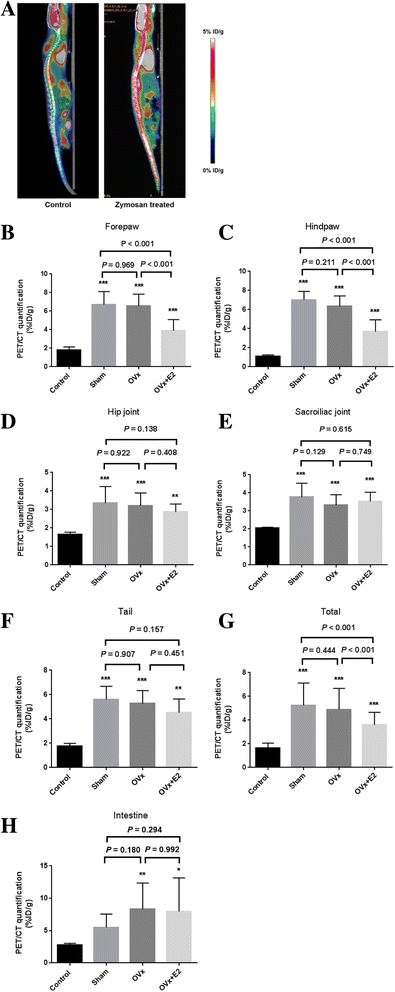
Quantitative measurement of 18F-fluorodeoxyglucose (18F-FDG) uptake. Visualization of 18F-FDG uptake in the spine of a wild-type (control) mouse and a mouse 8 weeks after zymosan injection (zymosan-treated) (a). Quantification of regions of interest (ROIs) in control, sham-operated (Sham), ovariectomy (OVx), and OVx + 17β-estradiol (OVx + E2) groups. b-g Forepaw (b), hind paw (c), hip joint (d), sacroiliac joint (e), tail (f), total of ROIs of five joints (g), and intestine (h). Values are mean ± SEM, n = 12 mice per group except for the wild-type control group (n = 2); *P < 0.05, **P < 0.01, ***P < 0.001 versus controls. PET/CT positron emission tomography/computed tomography
Fig. 3
Fig. 3
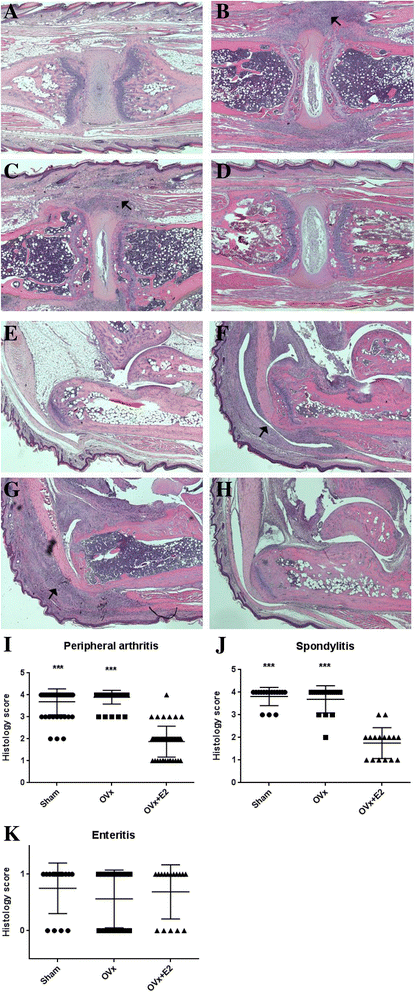
The effect of estrogen on microscopic inflammation. a-d The intervertebral area of the wild-type control (a), sham-operated (Sham) (b), ovariectomized (OVx) (c), and 17β-estradiol (E2)-treated (OVx + E2) mice (d). Arrows in b and c indicate inflammatory cell infiltration of the intervertebral disc area. e-f Achilles tendon of the normal control (e), sham (f), ovariectomized (g), and E2-treated mice (h). Arrows in f and g indicate inflammatory cell infiltration in the Achilles tendon. Inflammatory cell infiltration was not evident in E2-treated mice. Staining with H&E. Original magnification × 40. Histologic severity scores for peripheral joints affected by peripheral arthritis, including the forepaws, hind paws, ankle joints (i), tail (j), and intestine (k). Symbols represent individual mice, horizontal lines represent the mean, and whiskers represent the SEM; n = 16 mice per group except for the wild-type control (n = 2); *P < 0.05, **P < 0.01, ***P < 0.001 versus controls
Fig. 4
Fig. 4
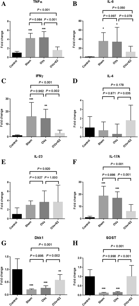
Local mRNA expression in the hind paws and forepaws was analyzed using the QuantiGene 2.0 Plex assay. Fold change in gene expression was compared to that in wild-type controls. a-h TNFɑ (a), IL-6 (b), interferon-γ (IFN-γ) (c), IL-4 (d), IL-23 (e), IL-17A (f), dickkopf-related protein 1 (Dkk1) (g), and sclerostin (SOST) (h). Data are expressed as the average fold change relative to the wild-type control group. Values are mean ± SEM; n = 12 mice per group except for the wild-type control (n = 2); *P < 0.05, **P < 0.01, ***P < 0.001, versus controls. Sham = sham-operated mice, OVx = ovariectomized mice, OVx + E2 = 17β-estradiol-treated ovariectomized mice

References

    1. Lee W, Reveille JD, Davis JC, Jr, Learch TJ, Ward MM, Weisman MH. Are there gender differences in severity of ankylosing spondylitis? Results from the PSOAS cohort. Ann Rheum Dis. 2007;66:633–8. doi: 10.1136/ard.2006.060293. - DOI - PMC - PubMed
    1. Rudwaleit M, van der Heijde D, Landewe R, Listing J, Akkoc N, Brandt J, et al. The development of Assessment of SpondyloArthritis international Society classification criteria for axial spondyloarthritis (part II): validation and final selection. Ann Rheum Dis. 2009;68:777–83. doi: 10.1136/ard.2009.108233. - DOI - PubMed
    1. Sieper J, van der Heijde D. Review: Nonradiographic axial spondyloarthritis: new definition of an old disease? Arthritis Rheum. 2013;65:543–51. doi: 10.1002/art.37803. - DOI - PubMed
    1. Oostveen J, Prevo R, den Boer J, van de Laar M. Early detection of sacroiliitis on magnetic resonance imaging and subsequent development of sacroiliitis on plain radiography. A prospective, longitudinal study. J Rheumatol. 1999;26:1953–8. - PubMed
    1. Rudwaleit M, Haibel H, Baraliakos X, Listing J, Marker-Hermann E, Zeidler H, et al. The early disease stage in axial spondylarthritis: results from the German Spondyloarthritis Inception Cohort. Arthritis Rheum. 2009;60:717–27. doi: 10.1002/art.24483. - DOI - PubMed

Publication types

MeSH terms

LinkOut - more resources