. 2018 Jan 1;314(1):L93-L106.
doi: 10.1152/ajplung.00091.2017. Epub 2017 Sep 7.

Opsin 3 and 4 mediate light-induced pulmonary vasorelaxation that is potentiated by G protein-coupled receptor kinase 2 inhibition

Affiliations

Opsin 3 and 4 mediate light-induced pulmonary vasorelaxation that is potentiated by G protein-coupled receptor kinase 2 inhibition

Sebastian Barreto Ortiz et al. Am J Physiol Lung Cell Mol Physiol. .

Abstract

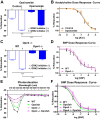
We recently demonstrated that blue light induces vasorelaxation in the systemic mouse circulation, a phenomenon mediated by the nonvisual G protein-coupled receptor melanopsin (Opsin 4; Opn4). Here we tested the hypothesis that nonvisual opsins mediate photorelaxation in the pulmonary circulation. We discovered Opsin 3 (Opn3), Opn4, and G protein-coupled receptor kinase 2 (GRK2) in rat pulmonary arteries (PAs) and in pulmonary arterial smooth muscle cells (PASMCs), where the opsins interact directly with GRK2, as demonstrated with a proximity ligation assay. Light elicited an intensity-dependent relaxation of PAs preconstricted with phenylephrine (PE), with a maximum response between 400 and 460 nm (blue light). Wavelength-specific photorelaxation was attenuated in PAs from Opn4-/- mice and further reduced following shRNA-mediated knockdown of Opn3. Inhibition of GRK2 amplified the response and prevented physiological desensitization to repeated light exposure. Blue light also prevented PE-induced constriction in isolated PAs, decreased basal tone, ablated PE-induced single-cell contraction of PASMCs, and reversed PE-induced depolarization in PASMCs when GRK2 was inhibited. The photorelaxation response was modulated by soluble guanylyl cyclase but not by protein kinase G or nitric oxide. Most importantly, blue light induced significant vasorelaxation of PAs from rats with chronic pulmonary hypertension and effectively lowered pulmonary arterial pressure in isolated intact perfused rat lungs subjected to acute hypoxia. These findings show that functional Opn3 and Opn4 in PAs represent an endogenous "optogenetic system" that mediates photorelaxation in the pulmonary vasculature. Phototherapy in conjunction with GRK2 inhibition could therefore provide an alternative treatment strategy for pulmonary vasoconstrictive disorders.

Keywords: GRK2; opsins; photorelaxation; pulmonary artery smooth muscle; pulmonary hypertension.


Figures

Fig. 1.
Fig. 1.
Expression of photorelaxation proteins in rat pulmonary arteries (PAs), rat pulmonary arterial smooth muscle cells (PASMCs), and human PASMCs. A: Opsin 3 (Opn3) and Opsin 4 (Opn4) were both detected in PAs, but Opsin 5 (Opn5) was not. B: G protein-coupled receptor kinase 2 (GRK2) was detected in rat PA (n = 5). Expression of Opn3, Opn4, and GRK2 in isolated rPASMCs via qRT-PCR (C) and Western blot (D) (n = 5) is shown. E: immunofluorescence images of hPASMCs stained for Opn3, Opn4, or GRK2 (n = 5). F and G: RT-PCR and qRT-PCR of hPASMC mRNA showing expression of Opn3, Opn4, and GRK2 but not Opn5 (n = 4–5). H: Western blot of hPASMC lysates tested for Opn3, Opn4, and GRK2 (n = 5) (+, with reverse transcriptase, RT; -, no RT). ***P < 0.001.
Fig. 2.
Fig. 2.
Vasoreactivity of rat pulmonary arteries (PAs) in response to light stimulation. A: photorelaxation was observed with cold white light in increasing intensities (40,000–2,000,000 lux units) with and without G protein-coupled receptor kinase 2 (GRK2) inhibitor (1 μM). There was a dose-dependent relaxation to light intensity. Photorelaxation was more profound in vessels pretreated with GRK2 inhibitor (n = 8). B: statistical comparison of photorelaxation to red, green, and blue light with and without GRK2 inhibitor (n = 6). C: representative myograph showing photorelaxation to red (620–750 nm, red arrow), green (495–570 nm, green arrow), and blue light (380–495 nm, blue arrow). Photorelaxation to light was first performed without GRK2 inhibitor followed by photorelaxation response in the presence of GRK2 inhibitor. PE, phenylephrine. D: photorelaxation to wavelength-specific blue light (455 nm) with and without GRK2 inhibitor (n = 11). E: photorelaxation to specific wavelength from 370 nm to 730 nm in 30-nm increments. Maximum response was observed between 400 and 460 nm (n = 3). F: representative myograph showing photorelaxation in wavelength-specific light stimulation (370–700 nm) in 30-nm increments. G: PE dose response (1 nM–10 μM) in PAs treated with blue light and GRK2 inhibitor showed an abrogated contractility compared with control vessels (n = 10). Representative myography graphs of PE dose response in PAs kept in the dark (H) or treated with blue light and GRK2 inhibitor (I) are shown. ***P < 0.001.
Fig. 3.
Fig. 3.
Role of Opsin 4 (Opn4) and Opsin 3 (Opn3) in photorelaxation of the mouse pulmonary artery (PA). A: photorelaxation was significantly attenuated in wild-type (WT) mouse PAs treated with Opn4 antagonist. Attenuated photorelaxation was observed in both the absence and presence of G protein-coupled receptor kinase 2 (GRK2) inhibitor (n = 10–16). B: acetylcholine dose response (1 nM–10 μM) showed a similar response in PAs in the presence and absence of Opn4 antagonists (n = 7). C: photorelaxation in PAs was significantly attenuated in vessels from Opn4−/− mice in both the absence and presence of GRK2 inhibitor (n = 22) (n = 10). D: sodium nitroprusside (SNP) response was similar in vessels from WT and Opn4 knockout (Opn4−/−) mice. E: photorelaxation response to specific wavelength from 370 nm to 730 nm in 30-nm increments in PAs from WT (control) and Opn4−/− mice and for those that were treated with Opn3 shRNA for 72 h. Maximum response was observed at 400–430 nm (400 nm: WT 66 ± 4.7%, Opn3−/− 50 ± 7.0%, Opn4−/− 33 ± 7.5%, Opn3−/−/Opn4−/− 22 ± 5.9%, P < 0.001; 430 nm: WT 76 ± 4.5%, Opn3−/− 57 ± 6.4%, Opn4−/− 34 ± 7.2%, Opn3−/−/Opn4−/− 18 ± 3.3%, P < 0.001), which was significantly different among all 4 groups (n = 9–11). F: vessel responses to SNP showed no significant differences among the vessels transduced with Opn3 shRNA adenovirus and/or with deficiency of Opn4 (n = 8–12). ***P < 0.001.
Fig. 4.
Fig. 4.
G protein-coupled receptor kinase 2 (GRK2) desensitizes the photorelaxation response and interacts directly with Opn3 and Opn4. A: repeated blue light (455 nm) stimulation on rat pulmonary arteries (PAs) with or without GRK2 inhibitor (n = 5). Attenuation in photorelaxation was observed in vessels not treated with GRK2 inhibitor, but no attenuation was observed in vessels treated with GRK2 inhibitor. B: representative myograph tracing showing repetitive blue light (blue arrows) response in rat PAs in the absence of GRK2 inhibitor followed by light response in the presence of GRK2 inhibitor. C: proximity ligation assay (PLA) of human pulmonary arterial smooth muscle cells (hPASMCs) tested for GRK2 and Opn3 or Opn4 proximity. Control stain was performed with only the GRK2 antibody (n = 5). D: PLA of hPASMCs tested for phosphoserine and Opn3 or Opn4 proximity. Control stain was performed with only the phosphoserine antibody. (n = 5). ***P < 0.001.
Fig. 5.
Fig. 5.
Blue light prevents phenylephrine (PE)-induced contractility in freshly isolated rat pulmonary arterial smooth muscle cells (rPASMCs) and causes hyperpolarization. Projected cell area (A) and individual cellular traction measurements (B) of freshly isolated rat PASMCs seeded on collagen-coated elastic gels embedded with either red fluorescent or yellow-green fluorescent beads and excited with green or blue light, respectively, are shown. C: individual cellular root mean square (RMS) traction measurements before and after addition of 10 μM PE (n = 20–35). D: Em measurements in primary rPASMCs in the presence and absence of G protein-coupled receptor kinase 2 (GRK2) inhibitor (1 μM). PASMCs were depolarized with either KCl (80 mM) or PE (10 μM) and then exposed to blue or red light (n = 5). E: diphosphorylation of myosin light chain (ppMLC) in rPASMCs was analyzed by immunoblotting of lysates from control cells without light exposure (lanes 1 and 2) and from cells exposed to blue light without (lanes 3 and 4) or after pretreatment with inhibitors for GRK2 (1 μM) (lanes 5 and 6) or Opn4 (opsinamide, 1 μM) (lanes 7 and 8). Densitometry analysis is shown normalized to control cells (n = 5). *P < 0.05, **P < 0.01, ***P < 0.001.
Fig. 6.
Fig. 6.
Photorelaxation in rat pulmonary arteries is nitric oxide (NO) independent. A: photorelaxation response in vessels treated and not treated with the NO synthase (NOS) inhibitor nitro-l-arginine methyl ester (l-NAME) (200 nM) showed no significant difference in maximum relaxation (n = 5–6). B: acetylcholine dose response (1 nM–10 μM) in vessels with and without l-NAME showed attenuated relaxation in vessels treated with l-NAME (n = 5–6). C: photorelaxation response in vessels treated and not treated with the NO scavenger cPTIO (300 μM) showed no difference in maximum relaxation (n = 5–6). D: acetylcholine dose response (1 nM–10 μM) in vessels treated and not treated with cPTIO showed attenuated relaxation in vessels treated with cPTIO (n = 5–6). ***P < 0.001.
Fig. 7.
Fig. 7.
Photorelaxation in rat pulmonary arteries involves sGC/cGMP. A: photorelaxation response in vessels treated and not treated with the sGC inhibitor 1H-[1,2,4]oxadiazolo[4,3-a]quinoxalin-1-one (ODQ) (0.5 μM) showed significant attenuation in vessels treated with ODQ (n = 5–6). B: sodium nitroprusside (SNP) dose response (1 nM–10 μM) in vessels treated and not treated with ODQ showed significant attenuation in relaxation in vessels treated with ODQ (n = 5–6). C: photorelaxation response in vessels treated and not treated with the sGC inhibitor NS2028 (0.1 μM) produced similar results (n = 12). ***P < 0.001.
Fig. 8.
Fig. 8.
Photorelaxation in rat pulmonary arteries is PKG independent. A: photorelaxation response in vessels treated and not treated with PKG inhibitor sc201161 showed no difference in relaxation (n = 7). B: acetylcholine dose response (1 nM–10 μM) in vessels treated and not treated with sc201161 showed significant attenuation in vessels treated with sc201161 (n = 5–6). C: photorelaxation response in vessels treated and not treated with PKG inhibitor Rp-8-Br-PET-cGMPS also showed no difference in relaxation (n = 6). D: acetylcholine dose response (1 nM–10 μM) in vessels treated and not treated with Rp-8-Br-PET-cGMPS showed significant attenuation in vessels treated with Rp-8-Br-PET-cGMPS (n = 6). **P < 0.01, ***P < 0.001.
Fig. 9.
Fig. 9.
Photorelaxation in disease rat models. A: photorelaxation with G protein-coupled receptor kinase 2 (GRK2) inhibition of rat pulmonary arteries (PAs) showed a significant vasorelaxation response both in normal rats and in rats subject to pulmonary hypertension after Sugen hypoxia (SuHx) treatment (n = 5). B: acetylcholine dose response (1 nM–10 μM) in SuHx PAs compared with normal vessels showed significant attenuation in SuHx vessels (n = 5). C: phenylephrine (PE)-induced constriction of PAs from SuHx rats was abolished in PAs treated with GRK2 inhibitor and exposed to blue light compared with SuHx PAs kept in the dark (n = 5). D: representative traces of lung perfusion experiments showing hypoxic pulmonary vasoconstriction (HPV). Lungs were pretreated with GRK2 inhibitor and then exposed to a hypoxic gas mixture by switching the ventilator to 4% O2, resulting in increased pulmonary arterial pressure (PPA). At peak HPV, lungs were exposed to blue light. E: statistical comparison of changes in PPA (as a percentage of the maximum HPV) induced after 2 min of blue light exposure in the lung perfusion model in the absence and presence of GRK2 inhibitor. In lungs treated with GRK2 inhibitor, stimulation with blue light decreased PPA to a greater extent (n = 5–7 per group). *P < 0.05, ***P < 0.001.
Fig. 10.
Fig. 10.
Suggested photorelaxation pathway in pulmonary arterial smooth muscle cells. G protein-coupled receptor kinase 2 (GRK2) desensitizes the activation of Opn3 and Opn4. sGC is activated downstream, leading to an increase in cGMP levels, which regulate CNG K+ channels, leading to hyperpolarization and further vasorelaxation.

References

    1. An SS, Mitzner W, Tang W-Y, Ahn K, Yoon AR, Huang J, Kilic O, Yong HM, Fahey JW, Kumar S, Biswal S, Holgate ST, Panettieri RA Jr, Solway J, Liggett SB. An inflammation-independent contraction mechanophenotype of airway smooth muscle in asthma. J Allergy Clin Immunol 138: 294–297.e4, 2016. doi:10.1016/j.jaci.2015.12.1315. - DOI - PMC - PubMed
    1. Barreto-Ortiz SF, Fradkin J, Eoh J, Trivero J, Davenport M, Ginn B, Mao H-Q, Gerecht S. Fabrication of 3-dimensional multicellular microvascular structures. FASEB J 29: 3302–3314, 2015. doi:10.1096/fj.14-263343. - DOI - PMC - PubMed
    1. Barreto-Ortiz SF, Zhang S, Davenport M, Fradkin J, Ginn B, Mao H-Q, Gerecht S. A novel in vitro model for microvasculature reveals regulation of circumferential ECM organization by curvature. PLoS One 8: e81061, 2013. doi:10.1371/journal.pone.0081061. - DOI - PMC - PubMed
    1. Batenburg WW, Kappers MH, Eikmann MJ, Ramzan SN, de Vries R, Danser AH. Light-induced vs. bradykinin-induced relaxation of coronary arteries: do S-nitrosothiols act as endothelium-derived hyperpolarizing factors? J Hypertens 27: 1631–1640, 2009. doi:10.1097/HJH.0b013e32832bff54. - DOI - PubMed
    1. Blasic JR Jr, Matos-Cruz V, Ujla D, Cameron EG, Hattar S, Halpern ME, Robinson PR. Identification of critical phosphorylation sites on the carboxy tail of melanopsin. Biochemistry 53: 2644–2649, 2014. doi:10.1021/bi401724r. - DOI - PMC - PubMed

Publication types

MeSH terms