Real-Time Motor Cortex Mapping for the Safe Resection of Glioma: An Intraoperative Resting-State fMRI Study
- PMID: 28882861
- PMCID: PMC7963570
- DOI: 10.3174/ajnr.A5369
Real-Time Motor Cortex Mapping for the Safe Resection of Glioma: An Intraoperative Resting-State fMRI Study
Abstract
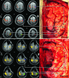
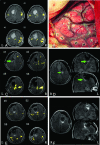
Background and purpose: Resting-state functional MR imaging has been used for motor mapping in presurgical planning but never used intraoperatively. This study aimed to investigate the feasibility of applying intraoperative resting-state functional MR imaging for the safe resection of gliomas using real-time motor cortex mapping during an operation.
Materials and methods: Using interventional MR imaging, we conducted preoperative and intraoperative resting-state intrinsic functional connectivity analyses of the motor cortex in 30 patients with brain tumors. Factors that may influence intraoperative imaging quality, including anesthesia type (general or awake anesthesia) and tumor cavity (filled with normal saline or not), were studied to investigate image quality. Additionally, direct cortical stimulation was used to validate the accuracy of intraoperative resting-state fMRI in mapping the motor cortex.
Results: Preoperative and intraoperative resting-state fMRI scans were acquired for all patients. Fourteen patients who successfully completed both sufficient intraoperative resting-state fMRI and direct cortical stimulation were used for further analysis of sensitivity and specificity. Compared with those subjected to direct cortical stimulation, the sensitivity and specificity of intraoperative resting-state fMRI in localizing the motor area were 61.7% and 93.7%, respectively. The image quality of intraoperative resting-state fMRI was better when the tumor cavity was filled with normal saline (P = .049). However, no significant difference between the anesthesia types was observed (P = .102).
Conclusions: This study demonstrates the feasibility of using intraoperative resting-state fMRI for real-time localization of functional areas during a neurologic operation. The findings suggest that using intraoperative resting-state fMRI can avoid the risk of intraoperative seizures due to direct cortical stimulation and may provide neurosurgeons with valuable information to facilitate the safe resection of gliomas.
© 2017 by American Journal of Neuroradiology.
Figures


References
-
- Krishnan R, Raabe A, Hattingen E, et al. . Functional magnetic resonance imaging-integrated neuronavigation: correlation between lesion-to-motor cortex distance and outcome. Neurosurgery 2004;55:904–14; discusssion 914–15 - PubMed
MeSH terms
LinkOut - more resources
Full Text Sources
Other Literature Sources
Medical
