. 2017 Oct 2;149(10):921-934.
doi: 10.1085/jgp.201711842. Epub 2017 Sep 7.

Slow fusion pore expansion creates a unique reaction chamber for co-packaged cargo

Affiliations

Slow fusion pore expansion creates a unique reaction chamber for co-packaged cargo

Kevin P Bohannon et al. J Gen Physiol. .

Abstract

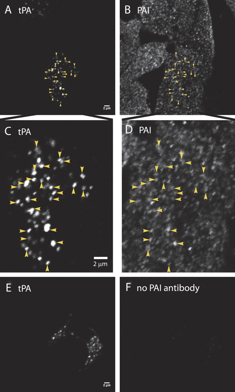
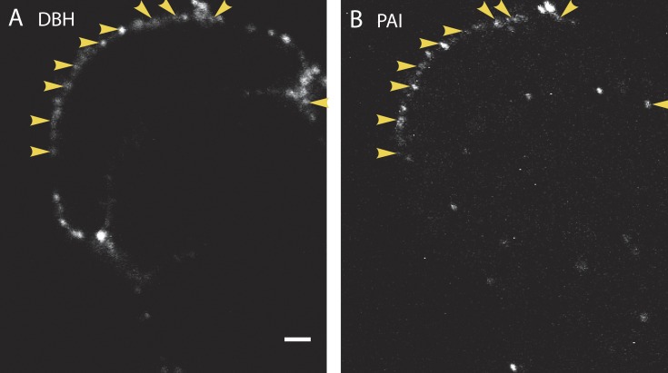
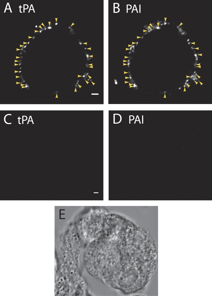
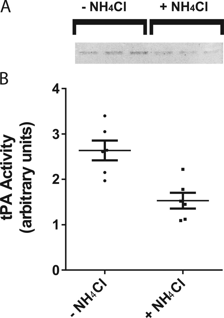
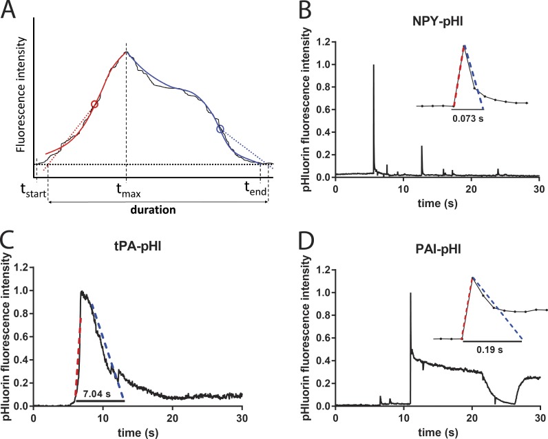
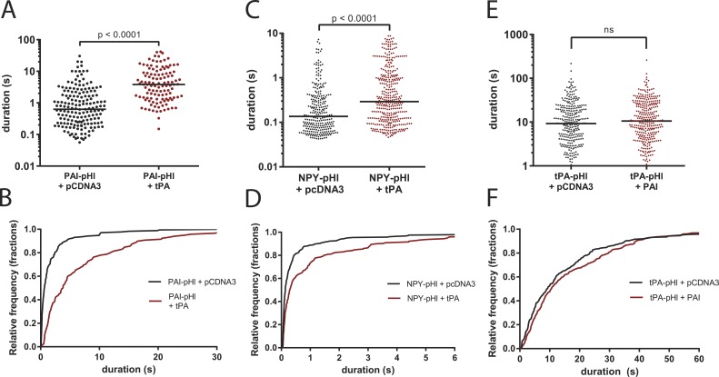
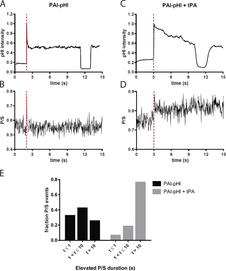
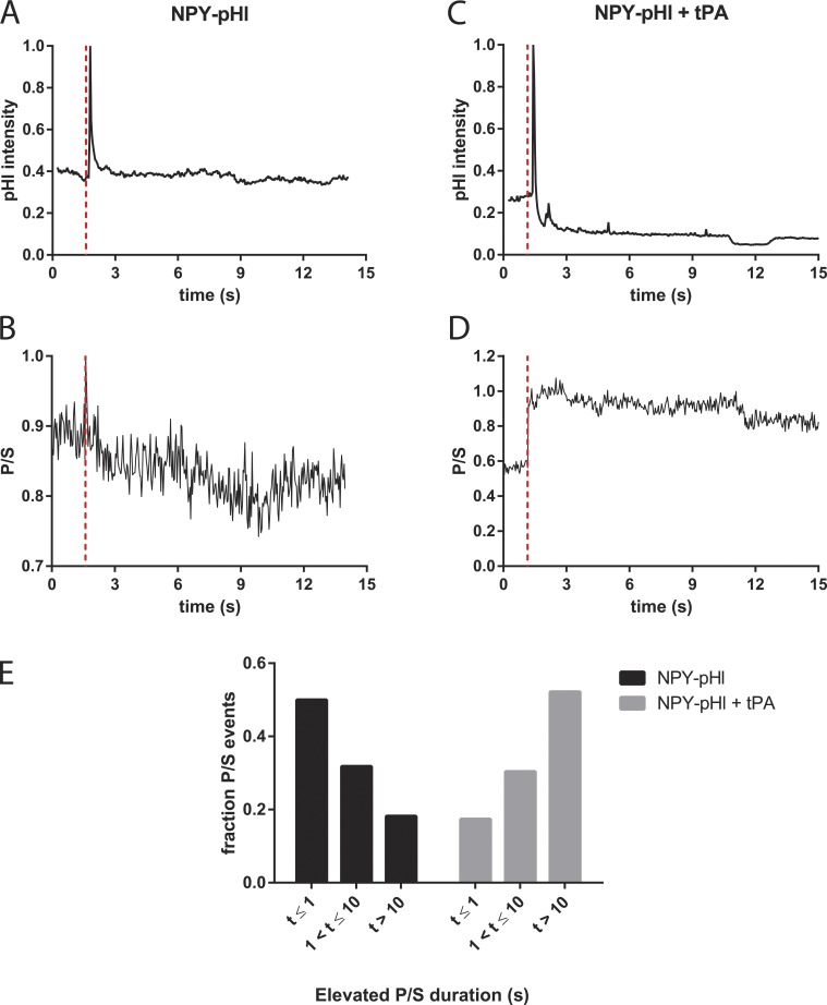
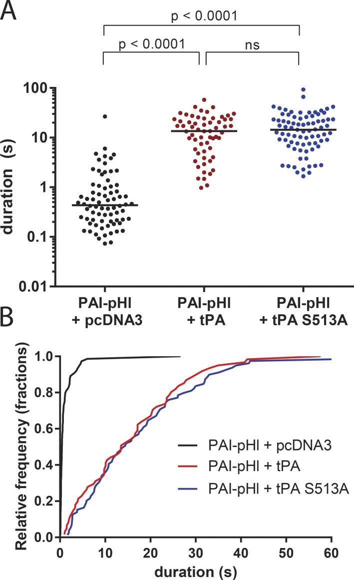
A lumenal secretory granule protein, tissue plasminogen activator (tPA), greatly slows fusion pore dilation and thereby slows its own discharge. We investigated another outcome of the long-lived narrow fusion pore: the creation of a nanoscale chemical reaction chamber for granule contents in which the pH is suddenly neutralized upon fusion. Bovine adrenal chromaffin cells endogenously express both tPA and its primary protein inhibitor, plasminogen activator inhibitor 1 (PAI). We found by immunocytochemistry that tPA and PAI are co-packaged in the same secretory granule. It is known that PAI irreversibly and covalently inactivates tPA at neutral pH. We demonstrate with zymography that the acidic granule lumen protects tPA from inactivation by PAI. Immunocytochemistry, total internal reflection fluorescence (TIRF) microscopy, and polarized TIRF microscopy demonstrated that co-packaged PAI and tPA remain together in granules for many seconds in the nanoscale reaction chamber, more than enough time to inhibit tPA and create a new secreted protein species.


Figures

Figure 1.
Figure 1.
PAI colocalizes with endogenous tPA in secretory granules. (A, C, and E) Cultured bovine chromaffin cells were fixed with 4% paraformaldehyde, permeabilized with methanol, and incubated with a primary antibody to tPA (rabbit anti-mouse tPA; Molecular Innovations), followed by Alexa Fluor 488–labeled goat anti–rabbit Fab fragments (Jackson ImmunoResearch Laboratories). Fab fragments rather than bivalent antibodies were used to preclude capture of a second rabbit primary antibody in a subsequent labeling step. After rinsing, the cells were blocked with an excess of unlabeled goat anti–rabbit Fab fragments (Jackson ImmunoResearch Laboratories), to ensure that none of the first primary ab (rabbit anti-tPA) would be accessible to a second anti-rabbit secondary antibody. (B, D, and F) Cells were next incubated with (B, D) or without (F) rabbit anti–human PAI (Abcam), followed by an Alexa Fluor 546–labeled anti-rabbit secondary antibody (B, D, and F; Molecular Probes). Cells were imaged by confocal microscopy. Images to be compared directly (e.g., A and E; B and F) were acquired at the same microscope settings, and the brightness and contrast were adjusted identically in making the figures. The absence of immunofluorescence in F (with no second primary antibody against PAI) indicates that the first rabbit primary antibody visualized in E was completely blocked before the addition of the second primary (seen in B and D). Colocalization of PAI (B) and tPA (A) is indicated by arrowheads and at an expanded scale in D and C, respectively. Bars, 2 µm.
Figure 2.
Figure 2.
Endogenous PAI colocalizes with DBH on the cell surface after stimulation. Cultured bovine chromaffin cells were stimulated for 10 s with 56 mM K+ at 34°C. The solution was replaced with buffer containing 5.6 mM K+, and the cells were immediately placed on ice. Cells were then incubated with antibodies to PAI (B) and to the lumenal domain of the granule membrane protein DBH (A) for 60 min on ice, and then processed and imaged by confocal microscopy. Because the cells were not permeabilized, only antigens present on the surface of the cells are visible. Arrowheads indicate instances of colocalization of secreted DBH and PAI. n = 10 cells. Bar, 2 µm.
Figure 3.
Figure 3.
Endogenous PAI colocalizes with tPA on the cell surface after stimulation. Cultured bovine chromaffin cells were incubated for 10 s in buffer with (A and B) or without (C–E) 56 mM K+ at 34°C. The solution was replaced with buffer containing 5.6 mM K+, and the cells were immediately placed on ice. Cells were then incubated with antibodies to tPA (A and C) and PAI (B and D) for 60 min on ice, and then processed and imaged by confocal microscopy. (A and B) Arrowheads indicate instances of colocalization of secreted tPA and PAI. When the fraction of puncta with colocalized tPA and PAI was calculated for n = 12 cells, it ranged from 67 to 90%, with a mean of 77.2 ± 2.2%. n = 333 total puncta. (C–E) Unstimulated cells, which were processed for tPA and PAI and visualized as in A and B, have little or no secreted tPA or PAI on the plasma membrane. Images that are to be compared directly (e.g., A and C; B and D) were acquired at the same microscope settings and adjusted to the same brightness and contrast when making the figures. Bars, 2 µm.
Figure 4.
Figure 4.
Neutralization of secretory granules allows inhibition of tPA activity. (A) Bovine chromaffin cells were incubated for 90 min in a physiological saline solution with or without 25 mM NH4Cl at 34°C to neutralize secretory granule pH. Cell lysates were resolved on a 10% SDS polyacrylamide gel containing casein (1 mg/ml) and plasminogen (10 µg/ml). SDS was removed by four washes in 2.5% Triton X-100 to allow renaturation of tPA. Gels (zymograms) were incubated in 100 mM Tris, pH 8.1, for 4 h at 37°C and then stained with Coomassie blue stain to visualize casein hydrolysis (inverted grayscale, in triplicate). (B) Gels were scanned, and band intensities were quantified in ImageJ. Mean ± SEM is shown. Student's t test resulted in a p-value of 0.0027.
Figure 5.
Figure 5.
NPY, tPA, and PAI have different secretion characteristics. Bovine chromaffin cells were transfected to express cargo proteins fused to pHl. Secretion was stimulated with 56 mM potassium buffer and observed by TIRF microscopy at a rate of 36 Hz. pHl fluorescence intensity was analyzed with a custom duration-finding program that is robust against variations in the shape of the data curve. (A) Schematic of program features. The solid thin black curve is the noisy fluorescence versus time of hypothetical data. A start time tstart is chosen, at which the fluorescence is defined to be baseline. Analysis is done separately for the rising phase (red) and the falling phase (blue), defined as before or after the fluorescence maximum time tmax, respectively. First, the fluorescence versus time in each phase is smoothed by fitting to a fifth-degree polynomial (thick red or blue solid lines). Next, a weighted average slope is calculated for each phase in the respective time windows (tstart, tmax) and (tmax, tend). Straight lines with those slopes (shown as dotted lines) are pinned to the maximum slope points (denoted by circles) and then extrapolated to the baseline to determine the event duration. (B) NPY-pHl is secreted rapidly. Entire events frequently take less than five frames at 36 Hz (inset; each point is one frame). Although the analysis was performed on a region of interest encompassing only the largest (first) fluorescent change, fluorescence changes from nearby fusion events are also evident. (C) tPA-pHl is secreted slowly, frequently lasting many seconds. (D) PAI-pHl is secreted rapidly. Often, ∼50% of PAI-pHl fluorescence is immediately lost, over just a few frames (inset). A fraction of PAI-pHl remains on the cell surface (plateau) and is sensitive to a pH 5.5 solution applied extracellularly.
Figure 6.
Figure 6.
tPA slows the release of co-packaged PAI-pHl and NPY-pHl. Bovine chromaffin cells were cotransfected to express cargo proteins tagged with pHl in tandem with tPA, PAI, or pCDNA3 vector control. Secretion was stimulated with 56 mM potassium buffer and observed by TIRF microscopy at a rate of 36 Hz. pHl fluorescence intensity was analyzed with a custom program as described in Fig. 5. (A and B) tPA slows PAI-pHl secretion. (C and D) tPA slows NPY-pHl secretion. (E and F) PAI does not slow the secretion of tPA-pHl. Each dot represents one fusion event, and medians are indicated by lines. Cumulative histograms plot data from upper panels. A Kolmogorov–Smirnoff test was performed for A and C. One-way ANOVA was performed in E with a post hoc Kruskal–Wallis test.
Figure 7.
Figure 7.
tPA slows fusion pore expansion in the presence of PAI-pHl. Bovine chromaffin cells were cotransfected with either PAI-pHl and pcDNA3 or PAI-pHl and tPA. pTIRF microscopy was performed as described in Materials and Methods. (A and C) Changes in pHluorin fluorescence were recorded over time. (B and D) Concurrently, DiD fluorescence as excited by P- and S-polarized light was recorded. The ratio P/S, plotted against time, corresponds to localized increases in membrane curvature. Secretion start time is indicated by the dotted red line. (E) Increases in P/S are reported semiquantitatively. The length of time P/S was elevated was measured for n = 40 PAI-pHl + pcDNA3 or n = 17 PAI-pHl = tPA events and binned as shown.
Figure 8.
Figure 8.
tPA slows fusion pore expansion in the presence of NPY-pHl. Bovine chromaffin cells were cotransfected with either NPY-pHl and pcDNA3 or NPY-pHl and tPA. pTIRF microscopy was performed as described in Materials and Methods. (A and C) Changes in pHl fluorescence were recorded over time. (B and D) Concurrently, DiD fluorescence as excited by P- and S-polarized light was recorded. The ratio P/S, plotted against time, corresponds to localized increases in membrane curvature. Secretion start time is indicated by the dotted red line. (E) Increases in P/S are reported semiquantitatively. The length of time P/S was elevated was measured for n = 22 NPY-pHl + pcDNA3 or n = 33 NPY-pHl = tPA events and binned as shown.
Figure 9.
Figure 9.
A kinase-dead mutant of tPA still slows PAI-pHl secretion. Bovine chromaffin cells were cotransfected to express PAI-pHl in tandem with tPA, S513A tPA, or empty vector control (pcDNA3). Secretion was stimulated with 56 mM potassium buffer and observed by TIRF microscopy at a rate of 36 Hz. pHl fluorescence intensity was analyzed with a custom program as described in Fig. 5. (A) Each dot represents one fusion event. Medians are indicated by lines. One-way ANOVA was performed with a post hoc Kruskal–Wallis test. (B) Cumulative histogram plots data from A.

Comment in

  • Chemistry in a vesicle.
    Jackson MB. Jackson MB. J Gen Physiol. 2017 Oct 2;149(10):893-896. doi: 10.1085/jgp.201711894. Epub 2017 Sep 12. J Gen Physiol. 2017. PMID: 28899933 Free PMC article.

References

    1. Anantharam A., Axelrod D., and Holz R.W.. 2010a Polarized TIRFM reveals changes in plasma membrane topology before and during granule fusion. Cell. Mol. Neurobiol. 30:1343–1349. 10.1007/s10571-010-9590-0 - DOI - PMC - PubMed
    1. Anantharam A., Onoa B., Edwards R.H., Holz R.W., and Axelrod D.. 2010b Localized topological changes of the plasma membrane upon exocytosis visualized by polarized TIRFM. J. Cell Biol. 188:415–428. 10.1083/jcb.200908010 - DOI - PMC - PubMed
    1. Anantharam A., Bittner M.A., Aikman R.L., Stuenkel E.L., Schmid S.L., Axelrod D., and Holz R.W.. 2011. A new role for the dynamin GTPase In the regulation of fusion pore expansion. Mol. Biol. Cell. 22:1907–1918. 10.1091/mbc.E11-02-0101 - DOI - PMC - PubMed
    1. Axelrod D. 1979. Carbocyanine dye orientation in red cell membrane studied by microscopic fluorescence polarization. Biophys. J. 26:557–573. 10.1016/S0006-3495(79)85271-6 - DOI - PMC - PubMed
    1. Bittner M.A., Aikman R.L., and Holz R.W.. 2013. A nibbling mechanism for clathrin-mediated retrieval of secretory granule membrane after exocytosis. J. Biol. Chem. 288:9177–9188. 10.1074/jbc.M113.450361 - DOI - PMC - PubMed

Substances

Associated data

LinkOut - more resources