. 2017 Nov 3;292(44):18372-18385.
doi: 10.1074/jbc.M117.801456. Epub 2017 Sep 7.

Kinetic signatures of myosin-5B, the motor involved in microvillus inclusion disease

Affiliations

Kinetic signatures of myosin-5B, the motor involved in microvillus inclusion disease

Sarah M Heissler et al. J Biol Chem. .

Abstract

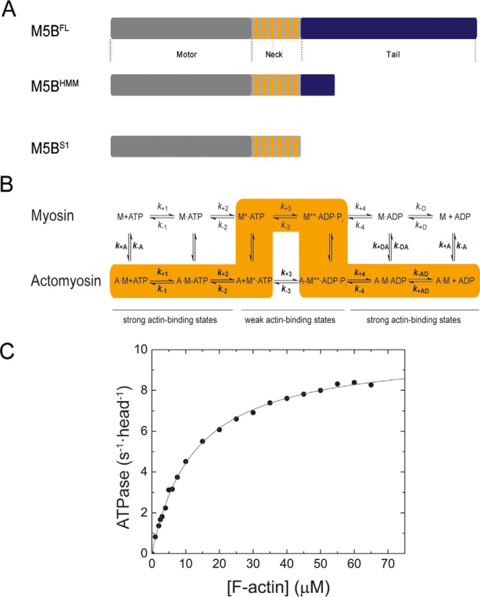
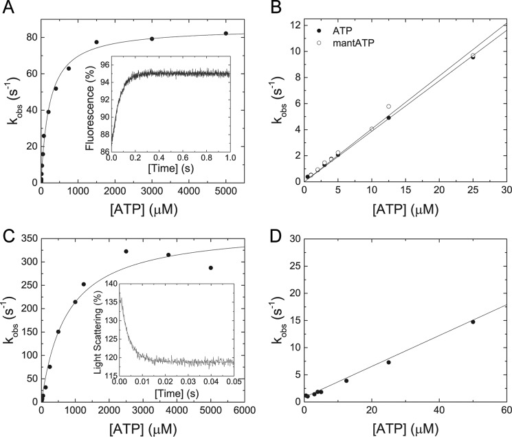
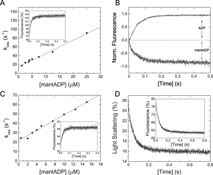
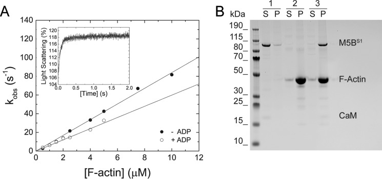
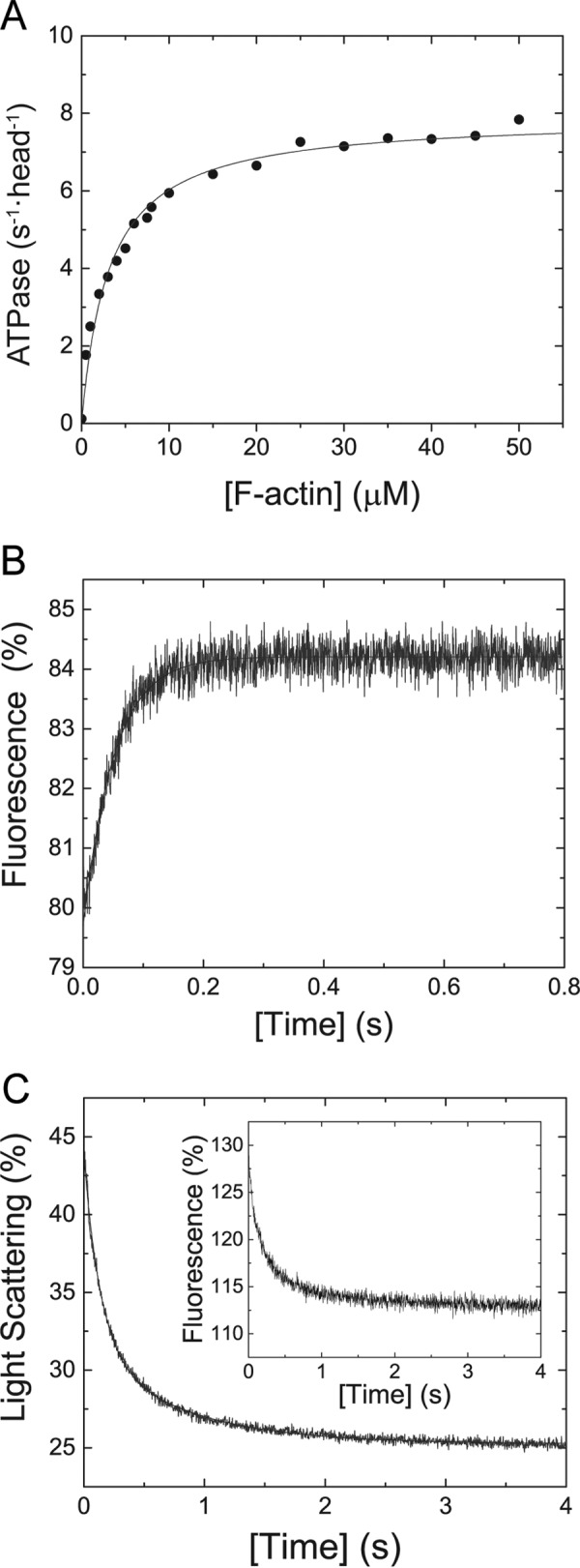
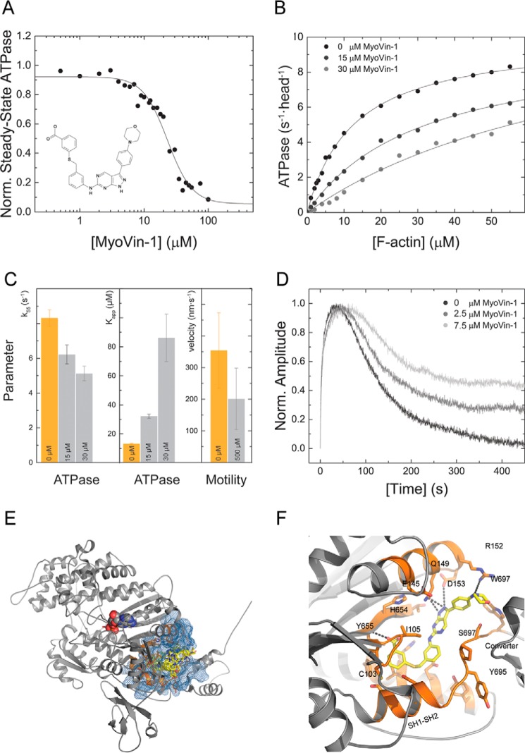
Myosin-5B is a ubiquitous molecular motor that transports cargo vesicles of the endomembrane system in intracellular recycling pathways. Myosin-5B malfunction causes the congenital enteropathy microvillus inclusion disease, underlining its importance in cellular homeostasis. Here we describe the interaction of myosin-5B with F-actin, nucleotides, and the pyrazolopyrimidine compound myoVin-1. We show that single-headed myosin-5B is an intermediate duty ratio motor with a kinetic ATPase cycle that is rate-limited by the release of phosphate. The presence of a second head generates strain and gating in the myosin-5B dimer that alters the kinetic signature by reducing the actin-activated ADP release rate to become rate-limiting. This kinetic transition into a high-duty ratio motor is a prerequisite for the proposed transport function of myosin-5B in cellular recycling pathways. Moreover, we show that the small molecule compound myoVin-1 inhibits the enzymatic and functional activity of myosin-5B in vitro Partial inhibition of the actin-activated steady-state ATPase activity and sliding velocity suggests that caution should be used when probing the effect of myoVin-1 on myosin-5-dependent transport processes in cells.

Keywords: ATPase; actin; inhibitor; kinetics; myoVin-1; myosin.


Conflict of interest statement

The authors declare that they have no conflicts of interest with the contents of this article

Figures

Figure 1.
Figure 1.
Domain organization, kinetic scheme, and steady-state ATPase activity of M5BS1. A, domain organization of myosin-5B and expression constructs used in this work. The myosin motor domain in the myosin heavy chain is shown in gray. The six IQ motifs in the myosin neck domain are shown in orange, and the tail domain is in blue. The tail domain mediates the dimerization of M5BFL and M5BHMM by means of a coiled-coil and interacts with binding partners. B, simplified kinetic scheme of the myosin and actomyosin ATPase cycle. The events of ATP binding, ATP hydrolysis, and phosphate release are shown for myosin in the actin detached (top row) and attached (bottom row) states. The main flux through the pathway is highlighted in orange, and the weak and strong actin-binding states are indicated. The notation distinguishes between the kinetic constants in the presence and absence of F-actin by using regular versus bold type; subscripts A and D refer to F-actin (KA) and ADP (KD), respectively. Dissociation equilibrium constants were calculated as Kx = k−x/k+x. M, myosin; A, actin. C, steady-state ATPase activity of M5BS1. Increasing concentrations of F-actin activate the steady-state ATPase activity to a kcat of 10.0 ± 0.1 s−1 with a Kapp of 12.6 ± 0.4 μm.
Figure 2.
Figure 2.
Transient kinetic interaction between MB5S1 and actoM5S1 with ATP and ATP analogs. A, the observed rate constant kobs depends hyperbolically on the ATP concentration and converges a value k+3 + k−3 of 85.4 ± 1.5 s−1. Half-saturation (1/K0.5) is reached at 225.1 + 14.7 μm ATP. Inset, the transient fluorescence increase represents the interaction between 50 μm ATP and 0.5 μm M5BS1 and has an observed rate constant kobs of 15.84 s−1. B, linear dependence of the observed rate constant kobs on [ATP] at low nucleotide concentrations. Binding of ATP or the nucleotide analog mantATP results in identical second-order rate constants of 0.39 ± 0.01 and 0.4 ± 0.01 μm−1 s−1, respectively. C, in the presence of F-actin, the observed rate constants kobs of the ATP-induced dissociation of the actomyosin complex show a hyperbolic dependence on the ATP concentration and converge to a value k+2 of 373.5 ± 18.7 s−1 with half-saturation (1/K0.5 = 1/K1) at 745.6 ± 120.7 μm ATP. Inset, time-dependent decrease in light scattering signal upon mixing 0.3 μm M5BS1 and 0.5 μm F-actin with 1250 μm ATP. The transient is best described with a single exponential fit with a kobs of 252.22 s−1. D, the observed rate constant kobs of the ATP-induced dissociation of the actomyosin complex linearly depends on the ATP concentration up to 50 μm substrate, defining K1k+2 to 0.28 ± 0.01 μm−1 s−1.
Figure 3.
Figure 3.
Transient kinetic interaction between M5S1 and actoM5S1 with ADP and ADP analogs. A, the observed rate constants upon rapidly mixing M5BS1 and mantADP follow a linear dependence in the concentration range tested. The straight line describes the second-order rate constant for ADP-binding k+D to 3.05 ± 0.16 μm−1 s−1. From the y intercept, the ADP release rate k−D was determined to be 14.3 ± 1.61 s−1. Inset, representative fluorescence increase after mixing 0.5 μm M5BS1 and 12.5 μm mantADP in a stopped-flow spectrophotometer. Single exponential fit to the transient results in a kobs of 48.65 s−1. B, ADP release kinetics from M5BS1. Shown are the normalized data of the time-dependent increase in the intrinsic fluorescence signal after mixing 0.5 μm M5BS1 in the presence of a 25 μm ADP with 1.5 mm ATP. The transient was fit to a single exponential, yielding the ADP release rate constant k−D = 18.9 s−1. Similar, rapidly mixing 0.5 μm M5BS1 in the presence of a 50-fold molar excess of ADP (5 μm mantADP and 20 μm ADP) with 1.5 mm ATP resulted in a k−D of 18.0 s−1. C, mantADP dependence of the observed rate constant kobs after mixing with actoM5BS1 under pseudo-first order conditions. The solid line describes the second-order binding rate constant k+AD = 2.71 ± 0.11 μm−1 s−1 and the ADP release rate constant k−AD = 21.9 ± 0.9 s−1. Inset, time-dependent fluorescence increase after mixing 0.5 μm actoM5BS1 with 7.5 μm mantADP. Single exponential fit yields a kobs = 42.39 s−1. D, dissociation of 0.5 μm actomyosin in the presence of 40 μm ADP with 2 mm ATP. A single exponential fit to the time course of the light scattering signal yields a k−AD of 17.13 s−1. Inset, representative transient of the interaction between 0.5 μm actomyosin and 40 μm ADP (2.5 μm mantADP and 37.5 μm ADP) with 2 mm ATP. A k−AD of 16.69 s−1 was determined from a single exponential fit to the data.
Figure 4.
Figure 4.
Transient kinetic interaction between M5BS1 and F-actin in the presence and absence of ADP. A, increasing concentrations of F-actin were mixed with M5BS1 in a stopped-flow spectrophotometer in the presence or absence of saturating ADP. Linear fits to the data sets describe the second-order binding rate constants k+A and k+DA to 8.39 ± 0.16 and 5.98 ± 0.19 μm−1 s−1, respectively. Inset, time-dependent change in light scattering after rapidly mixing 0.5 μm M5BS1 with 4 μm F-actin in the presence of 80 μm ADP. Single exponential fit to the data set results in a kobs of 13.61 s−1. B, a cosedimentation assay shows that an actomyosin complex is formed in SF buffer. Sample 1, 2 μm M5BS1 (control); sample 2, 10 μm F-actin (control); sample 3, 2 μm M5BS1 and 10 μm F-actin. Abbreviations S and P refer to supernatant and pellet, respectively.
Figure 5.
Figure 5.
Steady-state ATPase activity and key kinetic parameters of M5BHMM. A, the Michaelis–Menten parameters kcat = 7.91 ± 0.15 s−1 and Kapp = 3.12 ± 0.26 μm for M5BHMM were determined under steady-state conditions for M5BHMM. B, the transient fluorescence increase shows the interaction of 0.25 μm M5BHMM in the presence of 80 μm ADP after rapid mixing with 2 mm ATP. Single exponential fit to the data set yields a k−D,HMM of 19.53 s−1. C, dissociation of the actoM5BHMM·ADP complex (0.25 μm M5BHMM, 0.5 μm F-actin, 40 μm ADP) with 2 mm ATP results in a double-exponential decrease in light scattering signal (k−AD,HMM,fast = 7.24 s−1, k−AD,HMM,slow = 1.27 s−1). The fast phase has an amplitude of −12.4%, the slow phase an amplitude of −6.19%. Inset, similar ADP release rates and amplitudes (k−AD,HMM,fast = 7.1 s−1, k−AD,HMM,slow = 1.1 s−1, Afast = −10.67%, Aslow = −4.4%) are obtained after rapidly mixing the actoM5BHMM·mADP complex (0.25 μm M5BHMM, 0.5 μm F-actin, 1.25 μm mADP, 18.75 μm ADP) with 1 mm ATP.
Figure 6.
Figure 6.
Chemical inhibition of M5BS1 with the small molecule myoVin-1. A, myoVin-1 inhibits the steady-state ATPase activity of M5BS1 at a fixed actin concentration of 30 μm in a dose-dependent manner with a KI of 23.12 ± 2.07 μm. The data are normalized to the uninhibited control. The structure of the pyrazolopyrimidine compound myoVin-1 is shown in the inset. B, myoVin-1 changes the kcat and the Kapp of the M5BS1 actin-activated ATPase activity in a dose-dependent manner. C, effect of myoVin-1 on the steady-state ATPase activity of at 55 μm F-actin (k55) and Kapp of M5BS1 and gliding velocity. Orange bars represent the uninhibited control (0 μm myoVin-1). For k55 and Kapp, light gray bars represent the measured parameters in the presence of 15 and 30 μm myoVin-1 as indicated. For the in vitro motility data, the light gray bar represents the actin gliding velocity in the presence of 500 μm myoVin-1 as indicated. D, single-turnover measurements (0.25 μm M5BS1 and 0.15 μm mantATP) in the presence of 0, 2.5, and 7.5 myoVin-1. The data show that myoVin-1 predominantly decreases the release of the mantATP hydrolysis products in a concentration-dependent manner. E, favorable binding pocket of myoVin-1 in the homology model of the human myosin-5B motor domain. The site is located at a distance of 17 Å to the nucleotide-binding pocket. The myosin-5B motor domain is shown in gray cartoon representation, the nucleotide is in spheres, and the predicted myoVin-1 binding pocket is in orange. The pharmacophore is highlighted in blue, and myoVin-1 is in yellow stick representation. The best pose has a binding energy of −10.5 kcal/mol. Of the 10 best scored poses, 8 ligands bind to the highlighted binding pocket and vary in their binding energy by only 1 kcal/mol. F, close-up view of the best binding conformation of myoVin-1 in the predicted binding pocket. The small molecule is coordinated by residues of the seven-stranded β-sheets of the transducer and hydrophobic residues including Trp-697 from the converter and hydrophobic methylene group of Arg-152. The pyrazolopyrimidine group of myoVin-1 is further stabilized by Gln-149 of the α-helix that connects the seven-stranded β-sheet transducer. Color coding is according to E.
Figure 7.
Figure 7.
Location and impact of microvillus inclusion disease causing mutations in the myosin-5B motor domain. A, mutations in the myosin motor domain and their functional impact score and impact on myosin motor function. The localization to structural elements in the myosin motor domain to either the active site or the actin-binding region is indicated when applicable. B, the homology model of the human myosin-5B motor domain (gray cartoon representation) shows the localization of the respective disease-associated mutation. The mutations are colored according to severity from yellow to orange according to A. The nucleotide is highlighted in gray spheres. Mutations G168R and R219H are in the active site, and C514R/R531W/P619L is in the actin-binding region. Mutations V108G, G316R, R401H, N456S, and R656C are predicted to have allosteric effects on myosin motor function, whereas G143R, G435(R/E), and R656C may interfere with protein folding and induce protein instability (26).

Similar articles

Cited by

References

    1. Gross S. P., Tuma M. C., Deacon S. W., Serpinskaya A. S., Reilein A. R., and Gelfand V. I. (2002) Interactions and regulation of molecular motors in Xenopus melanophores. J. Cell Biol. 156, 855–865 - PMC - PubMed
    1. Hammer J. A. 3rd, and Sellers J. R. (2011) Walking to work: roles for class V myosins as cargo transporters. Nat. Rev. Mol. Cell Biol. 13, 13–26 - PubMed
    1. Jacobs D. T., Weigert R., Grode K. D., Donaldson J. G., and Cheney R. E. (2009) Myosin Vc is a molecular motor that functions in secretory granule trafficking. Mol. Biol. Cell 20, 4471–4488 - PMC - PubMed
    1. Lapierre L. A., Kumar R., Hales C. M., Navarre J., Bhartur S. G., Burnette J. O., Provance D. W. Jr., Mercer J. A., Bähler M., and Goldenring J. R. (2001) Myosin vb is associated with plasma membrane recycling systems. Mol. Biol. Cell 12, 1843–1857 - PMC - PubMed
    1. Nascimento A. A., Roland J. T., and Gelfand V. I. (2003) Pigment cells: a model for the study of organelle transport. Annu. Rev. Cell Dev. Biol. 19, 469–491 - PubMed

Publication types

MeSH terms

Supplementary concepts

Associated data