. 2017 Nov;207(3):1041-1051.
doi: 10.1534/genetics.117.300260. Epub 2017 Sep 7.

Apico-basal Polarity Determinants Encoded by crumbs Genes Affect Ciliary Shaft Protein Composition, IFT Movement Dynamics, and Cilia Length

Affiliations

Apico-basal Polarity Determinants Encoded by crumbs Genes Affect Ciliary Shaft Protein Composition, IFT Movement Dynamics, and Cilia Length

Khodor Hazime et al. Genetics. 2017 Nov.

Erratum in

Abstract

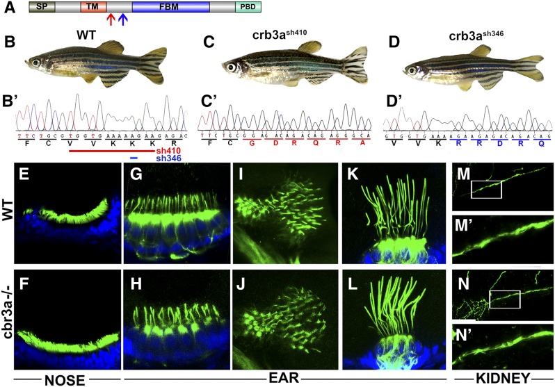
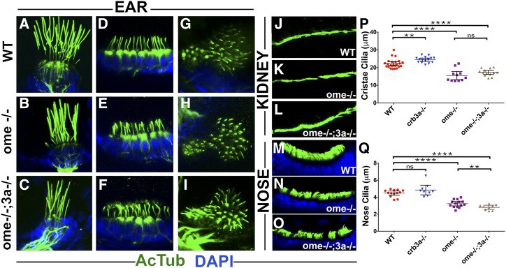
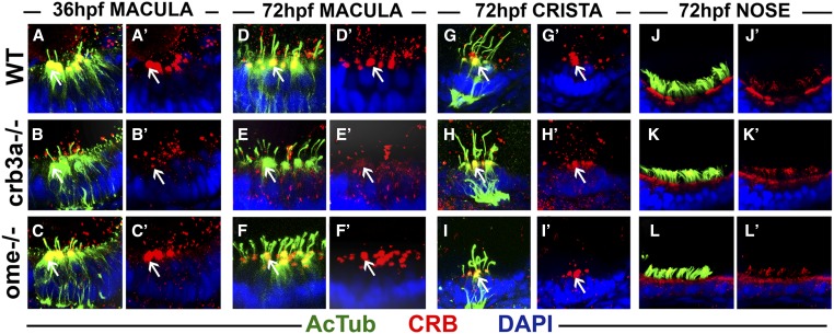
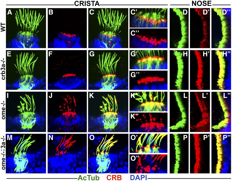
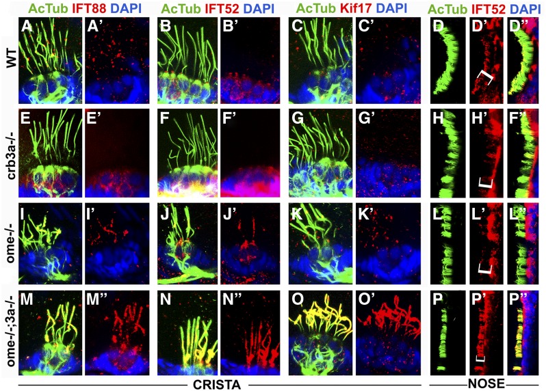
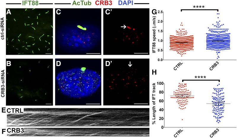
One of the most obvious manifestations of polarity in epithelia is the subdivision of the cell surface by cell junctions into apical and basolateral domains. crumbs genes are among key regulators of this form of polarity. Loss of crumbs function disrupts the apical cell junction belt and crumbs overexpression expands the apical membrane size. Crumbs proteins contain a single transmembrane domain and localize to cell junction area at the apical surface of epithelia. In some tissues, they are also found in cilia. To test their role in ciliogenesis, we investigated mutant phenotypes of zebrafish crumbs genes. In zebrafish, mutations of three crumbs genes, oko meduzy/crb2a, crb3a, and crb2b, affect cilia length in a subset of tissues. In oko meduzy (ome), this is accompanied by accumulation of other Crumbs proteins in the ciliary compartment. Moreover, intraflagellar transport (IFT) particle components accumulate in the ciliary shaft of ome;crb3a double mutants. Consistent with the above, Crb3 knockdown in mammalian cells affects the dynamics of IFT particle movement. These findings reveal crumbs-dependent mechanisms that regulate the localization of ciliary proteins, including Crumbs proteins themselves, and show that crumbs genes modulate intraflagellar transport and cilia elongation.

Keywords: Crumbs; IFT; apico-basal polarity; cilia; cristae.


Figures

Figure 1
Figure 1
crb3a mutant phenotype. (A) Schematic of Crb3a protein domain structure. Signal peptide (SP); transmembrane domain (TM); FERM-binding motif (FBM); and PDZ-binding domain (PBD) are indicated. Red arrow indicates the start of the frameshift in crb3a−/−sh410 mutant allele; blue arrow, the start of frameshift in crb3a−/−sh346 allele. (B–D) External phenotypes of wild-type (WT) (B), crb3a−/−sh410 homozygous mutant (C), and crb3a−/−sh346 homozygous mutant (D) adult zebrafish. (B’–D’) Sequences of wild-type (B’), and two mutant alleles: crb3a−/−sh410 (C’), and crb3a−/−sh346 (D’). Deletions in crb3a−/−sh410 (red line) and crb3a−/−sh346 (blue line) mutants are indicated in (B’). (E–N’) Images of wild-type and crb3a−/− mutant embryos stained with anti-acetylated tubulin antibody (in green) and counterstained with DAPI (in blue) at 5 days postfertilization: olfactory placode (E and F); anterior macula (G and H); posterior macula (I and J); lateral crista (K and L); and pronephros (M and N). (M’ and N’) are enlarged images of pronephric cilia shown in (M and N).
Figure 2
Figure 2
oko meduzy and crb3a genes modulate cilia length. (A–O) Whole-mount immunostaining of wild-type (WT), ome−/−, and ome−/−;3a−/− double mutant cilia in several tissues at 5 days postfertilization. (A–C) lateral crista, (D–F) anterior macula, (G–I) posterior macula, (J–L) pronephros, and (M–O) nasal pits. Zebrafish larvae were immunostained using anti-acetylated tubulin antibody (in green) and counterstained with DAPI (in blue) to visualize nuclei. (P) Graph of cilia length in the cristae of WT and crumbs mutants as indicated. Each dot represents the average length of all cilia in one crista. (Q) Graph of cilia length in the olfactory placodes of WT and crumbs mutants as indicated. In (P and Q), data were collected from at least two independent experiments using at least five animals per experiment. The mean and 95% C.I. are indicated. Based on Student’s t-tests; ** P < 0.01, *** P < 0.001, and **** P < 0.0001; not significant, ns. All differences were also significant based on Mann–Whitney test.
Figure 3
Figure 3
Crumbs expression in crb3a mutants at early stages of development. Confocal images of whole-mount cilia staining with anti-acetylated tubulin (AcTub) (green) and anti-CRB antibody (red). (A–C’) Staining of the otic vesicle at 36 h postfertilization (hpf). Crumbs proteins localize to the cilia base in the wild-type (WT) (A and A’) and ome−/− mutants (C and C’), but are absent in crb3a−/− mutant homozygotes (B and B’). At 72 hpf, Crumbs proteins still localize to the cilia base in maculae of WT animals (D and D’) and ome−/− mutants (F and F’), but very little signal is seen in crb3a−/− mutants (E and E’). No obvious differences are found in the localization of Crumbs proteins in the cristae of ome−/− (I and I’), crb3a−/− (H and H’), and WT individuals (G and G’). The localization patterns of Crumbs proteins in nasal pits of WT (J and J’), crb3a−/− mutant (K and K’), and ome−/− mutant (L and L’) animals do not show any obvious differences either. All samples were counterstained with DAPI to visualize nuclei. Arrows point to Crumbs signal at the apical surface of hair cells.
Figure 4
Figure 4
Crumbs proteins accumulate in cilia of ome mutants at 5 days postfertilization (dpf). Confocal images of whole-mount cilia staining with anti-acetylated tubulin antibody (AcTub) (green) and anti-CRB antibody (red) at 5 dpf. Shown are cristae and olfactory placodes of wild-types (WT), crb3a−/−, ome−/−, and ome−/−;crb3a−/− mutants as indicated. No obvious difference is seen in the localization of Crumbs proteins in cristae cilia of crb3a−/− mutants (E–G’’) when compared to WT (A–C’’). Crumbs proteins are strongly enriched in the cilia of nasal pits in crb3a−/− mutants (H–H’’) when compared to the WT (D–D’’). ome−/− and ome−/−;crb3a−/− double mutants show massive accumulation of Crumbs proteins inside cilia of cristae (I–K’’ and M–O’’) and nasal pits (L–L’’ and P–P’’). All samples were counterstained with DAPI to visualize nuclei (in blue).
Figure 5
Figure 5
Intraflagellar transport (IFT) particle components accumulate in the ciliary compartment of crumbs mutants. Confocal images of whole-mount cilia staining with antibodies to acetylated tubulin (AcTub) (green), and to IFT proteins (in red): IFT88, IFT52, and Kif17 at 5 days postfertilization. Shown are cristae and olfactory placodes of wild-type (WT), ome−/−, crb3a−/−, and ome−/−;crb3a−/− double mutants as indicated. IFT proteins are not detected in the ciliary shaft of WT cristae using this staining method (A–C’) and a low amount of IFT52 is found in WT olfactory cilia (D–D’’). Similarly, in crb3a−/− mutants, IFT proteins are not detected in cilia (E–H’’). Low levels of some IFT proteins are found in cristae cilia of ome−/− mutants (I–K’). Compared to WT, IFT52 localization is not affected in olfactory placode cilia of ome−/− mutants (L–L’’). In contrast to that, IFT proteins, including Kif17, strongly accumulate in cilia of ome−/−;crb3a−/− double mutants (M–P’’). All samples were counterstained with DAPI to visualize nuclei (in blue). Brackets in (D’, H’, L’, and P’) indicate nasal cilia.
Figure 6
Figure 6
CRB3 knockdown in mammalian cells affects intraflagellar transport (IFT) dynamics. (A and B) Maximum projections of total internal reflection fluorescence (TIRF) time-lapse recordings of IMCD3 cells grown on transwells and transfected with scrambled Ctrl-small interfering RNA (siRNA) (A) or CRB3-siRNA (B). These cells are stably transfected with an IFT88-GFP construct to visualize intraflagellar transport (green signal). (C–D’) Confocal images of control (CTRL) siRNA- (C and C’) and CRB3 siRNA-treated (D and D’) IMCD3 cells. Cilia are stained with antibodies to acetylated tubulin (AcTub) (green) and Crumbs (red). Samples are counterstained with DAPI to mark nuclei (in blue). (E and F) Kymographs of IFT movement in an IMCD3-IFT88 cell line transfected with CTRL or CRB3 siRNA as indicated. (G) Graph showing the speed of IFT particle movement in cilia of IFT88-GFP IMCD3 cells. Date collected from three independent experiments. (H) Lengths of IFT tracks in CTRL siRNA- and CRB3 siRNA-transfected cells expressed as percentages of total cilia length. Data are collected from two independent experiments. In (G and H), the mean and 95% C.I. are indicated. P < 10−4 based on Student’s t-tests. Bar, 10 μm (A and B) and 5 μm (C–D’).
Figure 7
Figure 7
crb2b affects cilia length. (A) Schematic of Crb2b protein domain structure (not to scale). Indicated are the signal peptide (SP), EGF-like repeats (E), Laminin G domains (L), transmembrane domain (TM), FERM-binding motif (FBM), and PDZ-binding domain (PBD). Red arrow shows the position of mutation in the crb2b−/−sa18042 mutant allele. (B and C) Phenotypes of wild-type (WT) (B) and crb2b−/−sa18042 (C) homozygous mutant adult zebrafish. (B’ and C’) Sequences of WT and crb2b−/−sa18042 mutant alleles. (D–I’) Whole-mount staining of WT and crb2b−/− mutants using anti-acetylated tubulin (AcTub) (green), and anti-CRB (red) antibodies at 5 days postfertilization. Samples were counterstained with DAPI to visualize nuclei (in blue). Crumbs proteins are not detected in the cilia of cristae (D–E’) and the lateral (LAT.) line (H–I’) of crb2b−/− mutants or their WT siblings. No differences in Crumbs signal are found between WT and mutants in the cilia of olfactory placodes (F–G’) and the pronephric duct (J–K’’). (L) Graph of cilia length in WT and crb2b−/− mutants. Each dot represents the average length of all cilia in one crista. Data were collected from three independent experiments using at least five animals per experiment. (M) Graph of cilia length in olfactory placodes of WT and crb2b−/− mutants. (N–O’) IFT proteins are not detected in the cristae cilia of WT (N and N’) and crb2b−/− mutants (O and O’). (P–Q’) IFT88 localization is not obviously different in nasal pit cilia of WT (P and P’) and crb2b−/− mutant (Q and Q’) animals. Brackets in (F, G, P, and Q) indicate nasal cilia. In (L and M), the mean and 95% C.I. are indicated. P < 0.001 based on Student’s t-test and Mann–Whitney test.

References

    1. Alves C. H., Bossers K., Vos R. M., Essing A. H. W., Swagemakers S., et al. , 2013. Microarray and morphological analysis of early postnatal CRB2 mutant retinas on a pure C57BL/6J genetic background. PLoS One 8: e82532. - PMC - PubMed
    1. Braasch I., Gehrke A. R., Smith J. J., Kawasaki K., Manousaki T., et al. , 2016. The spotted gar genome illuminates vertebrate evolution and facilitates human-teleost comparisons. Nat. Genet. 48: 427–437. - PMC - PubMed
    1. Chih B., Liu P., Chinn Y., Chalouni C., Komuves L. G., et al. , 2011. A ciliopathy complex at the transition zone protects the cilia as a privileged membrane domain. Nat. Cell Biol. 14: 61–72. - PubMed
    1. Craige B., Tsao C. C., Diener D. R., Hou Y., Lechtreck K. F., et al. , 2010. CEP290 tethers flagellar transition zone microtubules to the membrane and regulates flagellar protein content. J. Cell Biol. 190: 927–940. - PMC - PubMed
    1. den Hollander A. I., ten Brink J. B., de Kok Y. J., van Soest S., van den Born L. I., et al. , 1999. Mutations in a human homologue of Drosophila crumbs cause retinitis pigmentosa (RP12). Nat. Genet. 23: 217–221. - PubMed